Review
. 2018 Mar 2;17(1):101.
doi: 10.1186/s12936-018-2248-y.

Primaquine-induced haemolysis in females heterozygous for G6PD deficiency

Affiliations
Review

Primaquine-induced haemolysis in females heterozygous for G6PD deficiency

Cindy S Chu et al. Malar J. .

Abstract

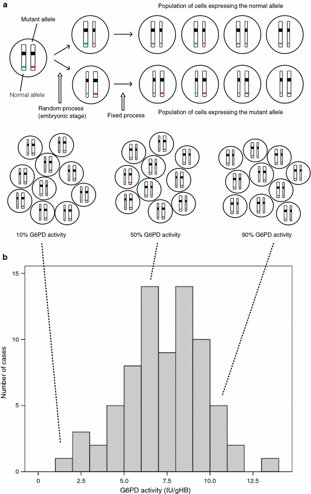
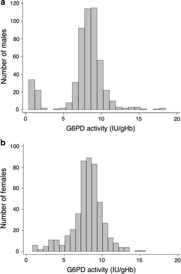
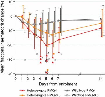
Oxidative agents can cause acute haemolytic anaemia in persons with G6PD deficiency. Understanding the relationship between G6PD genotype and the phenotypic expression of the enzyme deficiency is necessary so that severe haemolysis can be avoided. The patterns of oxidative haemolysis have been well described in G6PD deficient hemizygous males and homozygous females; and haemolysis in the proportionally more numerous heterozygous females has been documented mainly following consumption of fava beans and more recently dapsone. It has long been known that 8-aminoquinolines, notably primaquine and tafenoquine, cause acute haemolysis in G6PD deficiency. To support wider use of primaquine in Plasmodium vivax elimination, more data are needed on the haemolytic consequences of 8-aminoquinolines in G6PD heterozygous females. Two recent studies (in 2017) have provided precisely such data; and the need has emerged for the development of point of care quantitative testing of G6PD activity. Another priority is exploring alternative 8-aminoquinoline dosing regimens that are practical and improve safety in G6PD deficient individuals.

Keywords: 8-aminoquinoline; G6PD deficiency; G6PD heterozygous female; Haemolysis; Malaria; Plasmodium vivax; Primaquine; Radical cure.


Figures

Fig. 1
Fig. 1
Somatic cell mosaicism in G6PD heterozygous females and the associated G6PD activity (phenotype). X-chromosome inactivation and the phenotypic expression of G6PD deficiency in heterozygotes for GPPD mutations (a) (was Adapted from Baird et al. [61]). The top panel shows that at an early stage during embryonic development in each somatic cell of a female one of the two X chromosomes is inactivated (symbolized by a thin chromosome). In a heterozygote with one normal G6PD allele (blue) and one mutant (deficient) G6PD allele (red), after X-chromosome inactivation there are two types of cells: one type (top), where only the normal allele is expressed (blue stripe) will be G6PD normal; the other type (bottom) where only the mutant allele is expressed (red star), will be G6PD deficient. Once X inactivation has taken place it is faithfully maintained in the progeny of each cell. The bottom panel illustrates that, because X inactivation in the embryo is a random process, in adult tissue (e.g. red blood cells) the ratio between the number of cells in which one X-chromosome is inactive to the number of cells in which the other X-chromosome is active is variable: in these examples 1:9 (left), 5:5 (middle), 9:1 (right) (b) (was adapted from Bancone et al. [62]). This figure illustrates the distribution of G6PD activity in red cells from 74 G6PD heterozygous females. The G6PD activity is highly variable. The median activity is 11.76 IU/gHb so that 12 females, though heterozygous, are in the normal range, i.e. they appear to be G6PD normal (extreme phenotype). On the other hand, five females have ≲30% of the median activity, i.e. they are almost as G6PD deficient as a hemizygous male (extreme phenotype). The remaining females have intermediate G6PD levels. The dotted lines linking Fig. 1a to b show graphically how the extreme and intermediate red cell phenotypes arise
Fig. 2
Fig. 2
Phenotypic differences in quantitative G6PD activity between males and females. In a male population (a) there are two evident phenotypes (G6PD normal and deficient) as shown by the clearly bimodal distribution in the histogram. In a female population (b) some will have an intermediate phenotype as shown by the continuous distribution. (This figure was adapted from Oo et al. [26])
Fig. 3
Fig. 3
Mean fractional haematocrit changes over time in G6PD heterozygous and wild-type females taking primaquine. The line graph represents the fractional haematocrit plotted as the mean (95% CI). The plotted shapes represent individuals with maximum fractional haematocrit reductions below − 25%. The circled shapes represent individuals who received a blood transfusion. Het heterozygote, WT wild type, PMQ-1 primaquine dosed at 1 mg/kg/day for 7 days, PMQ-0.5 primaquine dosed at 0.5 mg/kg/day for 14 days. (This figure was taken from Chu et al. [49])

References

    1. Earle DP, Bigelow FS, Zubrod CG, Kane CA. Studies on the chemotherapy of the human malarias. IX. Effect of pamaquine on the blood cells of man. J Clin Invest. 1948;27:121–129. doi: 10.1172/JCI101950. - DOI - PMC - PubMed
    1. Alving AS, Pullman TN, Craige B, Jr, Jones R, Jr, Whorton CM, Eichelberger L. The clinical trial of 18 analogues of pamaquin (plasmochin) in vivax malaria Chesson strain. J Clin Invest. 1948;27:34–45. doi: 10.1172/JCI101963. - DOI - PubMed
    1. Maude RJ, Nguon C, Ly P, Bunkea T, Ngor P, de la Canavati Torre SE, et al. Spatial and temporal epidemiology of clinical malaria in Cambodia 2004–2013. Malar J. 2014;13:385. doi: 10.1186/1475-2875-13-385. - DOI - PMC - PubMed
    1. Betuela I, Rosanas-Urgell A, Kiniboro B, Stanisic DI, Samol L, de Lazzari E, et al. Relapses contribute significantly to the risk of Plasmodium vivax infection and disease in Papua New Guinean children 1–5 years of age. J Infect Dis. 2012;206:1771–1780. doi: 10.1093/infdis/jis580. - DOI - PubMed
    1. Sutanto I, Tjahjono B, Basri H, Taylor WRJ, Putri F, Meilia R, et al. Randomized, open-label trial of primaquine against vivax malaria relapse in Indonesia. Antimicrob Agents Chemother. 2013;57:1128–1135. doi: 10.1128/AAC.01879-12. - DOI - PMC - PubMed

MeSH terms