Review
. 2018 Mar 2;6(1):22.
doi: 10.1186/s40478-018-0515-3.

White matter changes in Alzheimer's disease: a focus on myelin and oligodendrocytes

Affiliations
Review

White matter changes in Alzheimer's disease: a focus on myelin and oligodendrocytes

Sara E Nasrabady et al. Acta Neuropathol Commun. .

Abstract

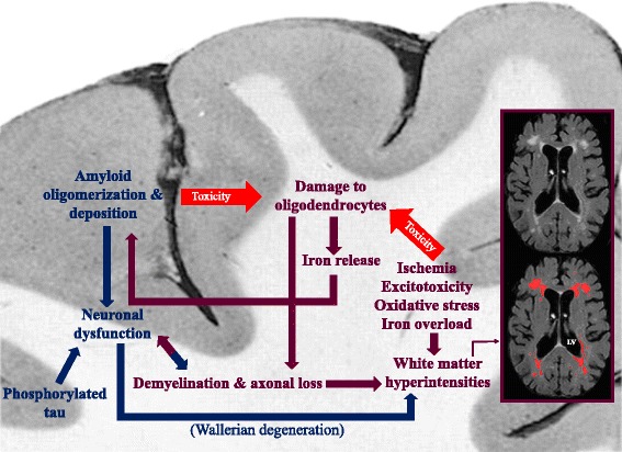
Alzheimer's disease (AD) is conceptualized as a progressive consequence of two hallmark pathological changes in grey matter: extracellular amyloid plaques and neurofibrillary tangles. However, over the past several years, neuroimaging studies have implicated micro- and macrostructural abnormalities in white matter in the risk and progression of AD, suggesting that in addition to the neuronal pathology characteristic of the disease, white matter degeneration and demyelination may be also important pathophysiological features. Here we review the evidence for white matter abnormalities in AD with a focus on myelin and oligodendrocytes, the only source of myelination in the central nervous system, and discuss the relationship between white matter changes and the hallmarks of Alzheimer's disease. We review several mechanisms such as ischemia, oxidative stress, excitotoxicity, iron overload, Aβ toxicity and tauopathy, which could affect oligodendrocytes. We conclude that white matter abnormalities, and in particular myelin and oligodendrocytes, could be mechanistically important in AD pathology and could be potential treatment targets.

Keywords: Alzheimer’s disease; Myelin, Oligodendrocyte; Neurodegeneration; Oxidative stress; White matter.


Conflict of interest statement

Competing interests

The authors declare that they have no competing interests.

Publisher’s Note

Springer Nature remains neutral with regard to jurisdictional claims in published maps and institutional affiliations.

Figures

Fig. 1
Fig. 1
This figure demonstrates examples of white matter myelin loss in brain sections from a patient with Alzheimer’s disease. Tissues from frontal and occipital areas are stained with Luxol fast blue-hematoxylin and eosin (LHE). b2 and b1 represent the regions with and without myelin pallor in the frontal area, respectively. d2 and d1 represent the regions with and without myelin pallor in the occipital area, respectively. The scale bars in images a, b, c and d are 1000 μm. The scale bars in images b1, b2, d1 and d2 are 100 μm
Fig. 2
Fig. 2
An example of Olig2+ oligodendrocyte distribution throughout the white matter from a neurologically-healthy adult, postmortem brain. The insets show Olig2+ nuclei at higher magnification. H&E counterstaining. Arrowheads: Olig2+ nuclei (brown). Dashed line: the border of white and grey matter. LV: lateral ventricle; SVWM: subventricular white matter; DWM: deep white matter; SCWM: subcortical white matter; GM: grey matter. The scale bars in a, b and c are 100 μm and the scale bars in the insets are 10 μm
Fig. 3
Fig. 3
This figure summarizes the pathological cascades, and their relation with each other, occurring during the development of Alzheimer’s disease in white matter and cortex. While ischemia, excitotoxicity, oxidative stress, and iron overload in white matter damage oligodendrocytes, on one hand, and amyloid toxicity affects them, on the other hand, the iron released from damaged oligodendrocytes promotes amyloid polymerization and deposition in grey matter. The consequent demyelination and axonal loss result in further white matter damage and neuronal dysfunction. Neuronal dysfunction is also a result of amyloid deposition in cortex and a proposed cause for white matter abnormalities in AD patients. White matter hyperintensities are labelled with red in the MRI (FLAIR) scan of an AD patient. Blue arrows: direction of the damages originating in grey matter. Maroon arrows: direction of the damages originating in white matter. LV lateral ventricle

References

    1. Al-Mashhadi S, Simpson JE, Heath PR, Dickman M, Forster G, Matthews FE, Brayne C, Ince PG, Wharton SB. Oxidative glial cell damage associated with white matter lesions in the aging human brain. Brain pathology (Zurich, Switzerland) 2015;25:565–574. doi: 10.1111/bpa.12216. - DOI - PMC - PubMed
    1. Back SA, Gan X, Li Y, Rosenberg PA, Volpe JJ. Maturation-dependent vulnerability of oligodendrocytes to oxidative stress-induced death caused by glutathione depletion. J Neurosci. 1998;18:6241–6253. - PMC - PubMed
    1. Back SA, Han BH, Luo NL, Chricton CA, Xanthoudakis S, Tam J, Arvin KL, Holtzman DM. Selective vulnerability of late oligodendrocyte progenitors to hypoxia-ischemia. J Neurosci. 2002;22:455–463. - PMC - PubMed
    1. Ballatore C, Lee VM, Trojanowski JQ. Tau-mediated neurodegeneration in Alzheimer's disease and related disorders. Nat Rev Neurosci. 2007;8:663–672. doi: 10.1038/nrn2194. - DOI - PubMed
    1. Bartzokis G (2011) Alzheimer’s disease as homeostatic responses to age-related myelin breakdown. Neurobiol Aging 32: 1341-1371 Doi 0.1016/j.Neurobiolaging.2009.08.007 - PMC - PubMed

Publication types