. 2018 Mar 2:10:376-386.
doi: 10.1016/j.omtn.2017.12.012. Epub 2017 Dec 30.

Gene Therapy via Trans-Splicing for LMNA-Related Congenital Muscular Dystrophy

Affiliations

Gene Therapy via Trans-Splicing for LMNA-Related Congenital Muscular Dystrophy

Feriel Azibani et al. Mol Ther Nucleic Acids. .

Abstract

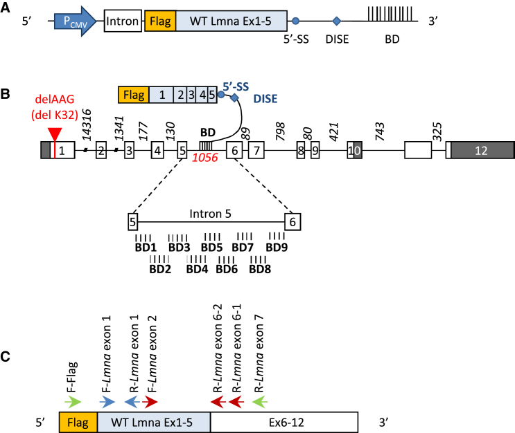
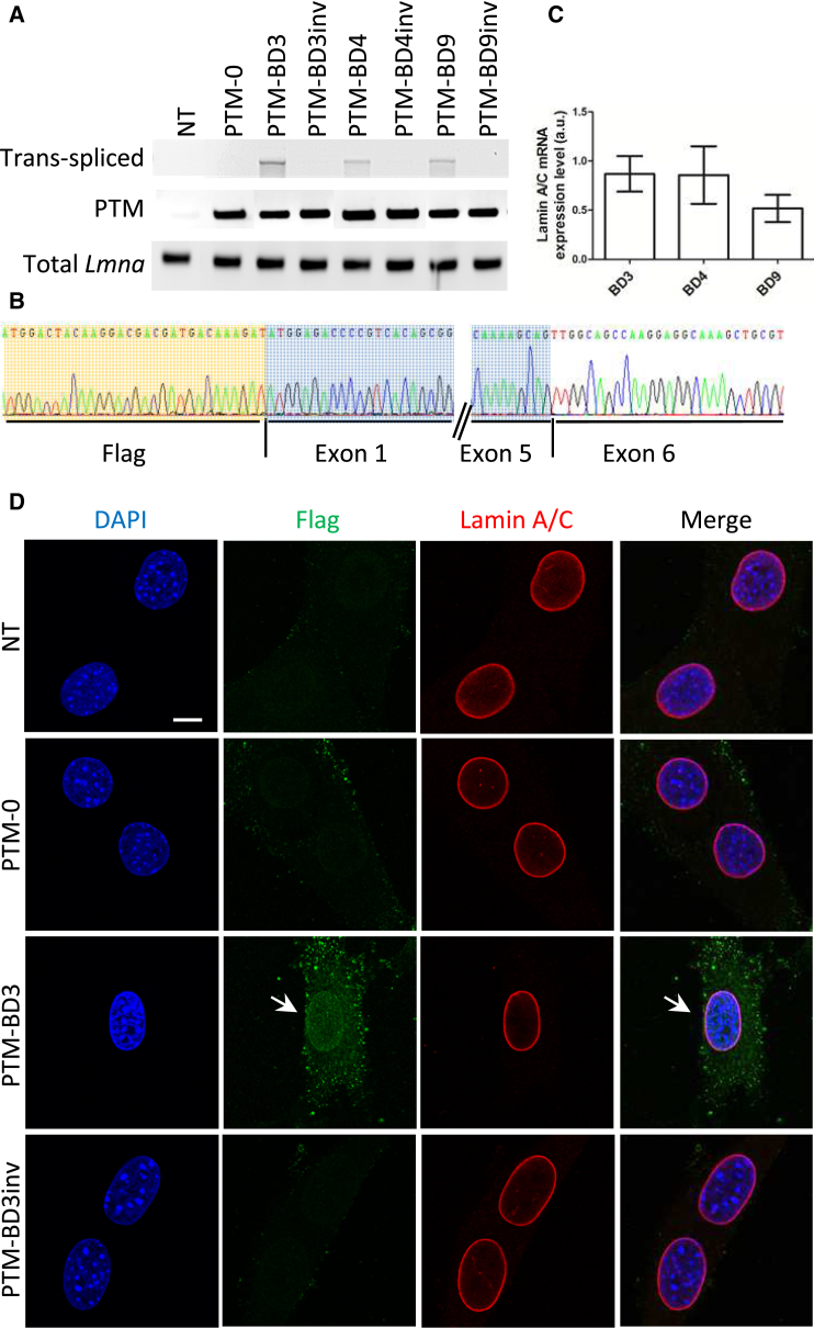
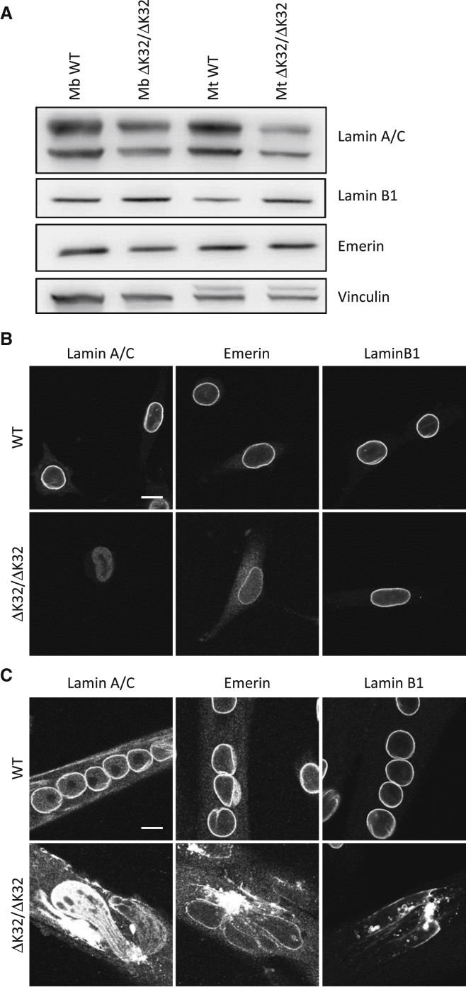
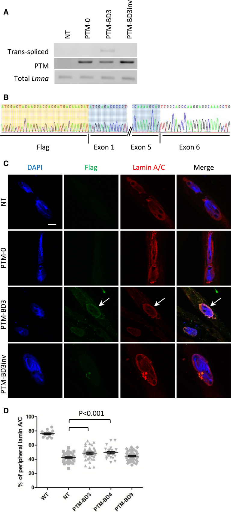
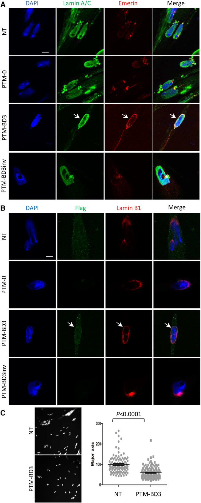
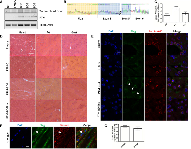
We assessed the potential of Lmna-mRNA repair by spliceosome-mediated RNA trans-splicing as a therapeutic approach for LMNA-related congenital muscular dystrophy. This gene therapy strategy leads to reduction of mutated transcript expression for the benefit of corresponding wild-type (WT) transcripts. We developed 5'-RNA pre-trans-splicing molecules containing the first five exons of Lmna and targeting intron 5 of Lmna pre-mRNA. Among nine pre-trans-splicing molecules, differing in the targeted sequence in intron 5 and tested in C2C12 myoblasts, three induced trans-splicing events on endogenous Lmna mRNA and confirmed at protein level. Further analyses performed in primary myotubes derived from an LMNA-related congenital muscular dystrophy (L-CMD) mouse model led to a partial rescue of the mutant phenotype. Finally, we tested this approach in vivo using adeno-associated virus (AAV) delivery in newborn mice and showed that trans-splicing events occurred in WT mice 50 days after AAV delivery, although at a low rate. Altogether, while these results provide the first evidence for reprogramming LMNA mRNA in vitro, strategies to improve the rate of trans-splicing events still need to be developed for efficient application of this therapeutic approach in vivo.

Keywords: LMNA-related congenital muscular dystrophy; Lmna; gene therapy; lamin A/C; trans-splicing.


Figures

Figure 1
Figure 1
5′ Trans-Splicing Strategy for Lmna Transcripts (A) Schematic illustration of part of the vector encoding for a pre-trans-splicing molecule (PTM). The PTM is a transcript comprising the FLAG coding sequence followed by the first five Lmna exons (WT Lmna ex 1–5), a 5′ splice site (5′SS) followed by a downstream intronic splicing enhancer (DISE), and a binding domain (BD) of 150 bp complementary to Lmna intron 5. PTM sequences are placed under control of a CMV promoter (PCMV). An intronic sequence was added to stabilize the transcript. (B) Schematic representation of Lmna pre-mRNA and PTMs. The mouse Lmna pre-messenger consists of exons (boxes) and introns (lines). White boxes represent coding sequences, and gray boxes represent non-coding sequence for prelamin A. Intronic size lengths are indicated. The red arrow refers to localization of the ΔK32 mutation in exon 1. Below, an enlarged view of intron 5 shows the localization of the different designed binding domains. (C) Schematic representation of a trans-spliced mRNA molecule and the different primers used to characterize trans-spliced/repaired mRNA (F-FLAG and R-Lmna exon 7), PTMs (F-FLAG and R-Lmna exon 1), and total (endogenous and trans-spliced) Lmna molecules (F-Lmna exon 1 and R-Lmna exon 1). Primers used for nested qPCR of trans-spliced Lmna transcripts are F-FLAG and R-Lmna exon 6-1 for the first PCR, followed by F-Lmna exon 2 and R-Lmna exon 6-2 for the qPCR.
Figure 2
Figure 2
Evaluation of PTM’s Efficacy in C2C12 Cells (A) PCR analysis was performed with primers localized on Lmna mRNA (Figure 1C), on RNA extracts from C2C12 myoblasts that were either non-transfected (NT) or transfected for 48 hr with PTM-0, PTM-BD, and PTM-BDinv plasmids. (B) Sequencing of trans-spliced Lmna amplicons from PTM-BD3-transfected cells after subcloning confirmed trans-splicing events with the junction between the exogenous FLAG-Lmna exons 1–5 (highlighted in orange and blue) with endogenous Lmna exon 6 and the following exons. (C) Bar graph showing the level of lamin A/C mRNA expression by nested qPCR. BD3, transfected with pSMD2-PTM-BD3 (n = 4); BD4, transfected with pSMD2-PTM-BD4 (n = 3); BD9, transfected with pSMD2-PTM-BD9 (n = 5). Differences are not statistically significant. Error bars correspond to SEM. (D) Immunofluorescence analysis of NT or transfected C2C12 with PTM-0, PTM-BD3, PTM-BD3inv. Cells were double-stained with anti-FLAG (green) and anti-Lamin A/C (red) antibodies. Nuclei were stained with DAPI (blue). Arrows show a positive FLAG-tagged nucleus. Scale bar: 10 μm.
Figure 3
Figure 3
Decreased Expression and Abnormal Localization of Δ32-Lamin A/C in Mouse Mutant Muscle Cells (A) Representative western blot analysis for mouse primary WT and mutant (ΔK32/ΔK32) myoblasts (Mb) and myotubes (Mt; 24 hr of differentiation) probed for lamin A/C, lamin B1, and emerin proteins relative to vinculin. (B and C) Immunofluorescent confocal micrographs of WT and mutant myoblasts (B) and 24-hr differentiated myotubes (C), stained for lamin A/C, emerin, and lamin B1. Scale bars: 10 μm.
Figure 4
Figure 4
Detection of Repaired Lmna mRNA Induced by 5′ Trans-Splicing in Mouse Mutant Primary Myotubes (A) PCR analysis was performed on RNA extracts of mouse primary mutant myotubes transduced or not (NT) for 7 days with AAV2/9 expressing PTM-0, PTM-BD3, or PTM-BD3inv using primers described in Figure 1C and Table S2. (B) Sequencing of the F-FLAG/R-Lmna exon 7 cloned amplicons from AAV2/9-PTM-BD3-transduced cells confirmed trans-splicing events with the junction between the exogenous FLAG-Lmna exons 1–5 (highlighted in orange and blue) and endogenous Lmna exon 6 and the following exons. (C) Immunofluorescence analysis of mutant myotubes that were non-transduced (NT) or transduced with AAV2/9-PTM-0, -PTM-BD3, and -PTM-BD3inv. Cells were double-stained with anti-FLAG (green) and anti-Lamin A/C (red) antibodies. Nuclei were stained with DAPI (blue). Arrows show a FLAG+ nucleus. Scale bar: 10 μm. (D) Scatterplot representing the percentage of lamin A/C staining found at the nuclear periphery in wild-type (WT; n = 19) and non-transduced (NT; n = 40) or PTM-BD3- (n = 40), PTM-BD4- (n = 24), or PTM-BD9 (n = 41)-transduced mutant myotubes. All groups are statistically different from WT (p < 0.001), while only PTM-BD3 and PTM-BD4 are statistically different from NT (p < 0.01). Error bars correspond to SEM.
Figure 5
Figure 5
5′ Trans-Splicing of Lmna Improves the Nuclear Phenotype of Mutant Myotubes (A and B) Immunofluorescence analysis of mutant myotubes that were non-transduced (NT) or transduced with AAV2/9-PTM-0, -PTM-BD3, and -PTM-BD3inv. Cells were differentiated and transduced for 7 days and then stained with lamin A/C (green)/emerin (red) (A) or FLAG (green)/Lamin B1 (red) (B). Nuclei were stained with DAPI (blue). Arrows point to repaired nuclei. Scale bars: 10 μm. (C) Representative confocal images of DAPI-stained myotubes that were either non-transduced (NT) or transduced with AAV2/9-PTM-BD3, and scatterplot showing the distribution and the mean size of nuclei long axis (NT: n = 107; PTM-BD3: n = 137). Error bars correspond to SEM.
Figure 6
Figure 6
Evidence for Reprogramming Lmna mRNA and Protein by 5′ Trans-Splicing In Vivo (A) PCR analysis was performed on RNA extracts of heart tissue from WT mice 50 days after systemic injection of AAV2/9-empty or expressing PTM-0, PTM-BD4, or PTM-BD4inv using primers described in Figure 1C and Table S2. (B) Sequencing of F-FLAG/R-Lmna exon 7 cloned amplicons from heart from mice 50 days after systemic injection with AAV2/9-PTM-BD4 confirmed trans-splicing events with the junction between the exogenous FLAG-Lmna exons 1–5 (highlighted in orange and blue) and endogenous Lmna exon 6. (C) Bar graph showing the level of lamin A/C mRNA expression by nested qPCR in hearts of WT mice 50 days after systemic injection of AAV2/9-PTM-BD3, -BD4, or -BD9. n = 4 per group, and differences are not statistically significant. Error bars correspond to SEM. (D) H&E staining of striated muscle sections from WT mice 50 days after systemic injection with AAV2/9-empty or expressing PTM-0, PTM-BD4 or PTM-BD4inv. Scale bar: 50 μm. (E) Immunofluorescence analysis of heart sections from WT mice 50 days after systemic injection of AAV2/9-PTM-0, -PTM-BD4, and -PTM-BD4inv. Cells were double-stained with anti-FLAG (green) and anti-Lamin A/C (red) antibodies. Nuclei were stained with DAPI (blue). Scale bar: 10 μm. (F) Immunofluorescence analysis of heart sections from WT mouse 50 days after systemic injection of AAV2/9-PTM-BD4. Cells were double-stained with anti-FLAG (green) and anti-desmin (red) antibodies. Nuclei were stained with DAPI (blue). Arrows point to a positive FLAG-tagged nucleus. Scale bar: 10 μm. (G) Bar graph showing the level of lamin A/C mRNA expression by nested qPCR in hearts of mice 15 (n = 3) or 50 days (n = 4) after systemic injection of AAV2/9-PTM-BD4. Differences are not statistically significant. Error bars correspond to SEM. Gast, gastrocnemius; TA, tibialis anterior.

References

    1. Broers J.L., Ramaekers F.C., Bonne G., Yaou R.B., Hutchison C.J. Nuclear lamins: laminopathies and their role in premature ageing. Physiol. Rev. 2006;86:967–1008. - PubMed
    1. Aebi U., Cohn J., Buhle L., Gerace L. The nuclear lamina is a meshwork of intermediate-type filaments. Nature. 1986;323:560–564. - PubMed
    1. Hozák P., Sasseville A.M., Raymond Y., Cook P.R. Lamin proteins form an internal nucleoskeleton as well as a peripheral lamina in human cells. J. Cell Sci. 1995;108:635–644. - PubMed
    1. Gruenbaum Y., Foisner R. Lamins: nuclear intermediate filament proteins with fundamental functions in nuclear mechanics and genome regulation. Annu. Rev. Biochem. 2015;84:131–164. - PubMed
    1. Bonne G., Di Barletta M.R., Varnous S., Bécane H.M., Hammouda E.H., Merlini L., Muntoni F., Greenberg C.R., Gary F., Urtizberea J.A. Mutations in the gene encoding lamin A/C cause autosomal dominant Emery-Dreifuss muscular dystrophy. Nat. Genet. 1999;21:285–288. - PubMed

LinkOut - more resources