. 2018 Apr 15;200(8):3000-3007.
doi: 10.4049/jimmunol.1700407. Epub 2018 Mar 2.

Functional Anti-TIGIT Antibodies Regulate Development of Autoimmunity and Antitumor Immunity

Affiliations

Functional Anti-TIGIT Antibodies Regulate Development of Autoimmunity and Antitumor Immunity

Karen O Dixon et al. J Immunol. .

Abstract

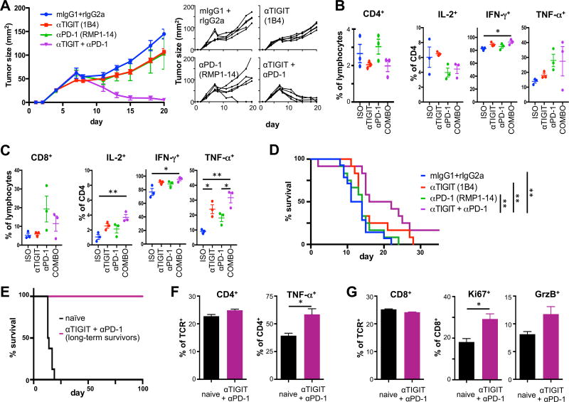
Coinhibitory receptors, such as CTLA-4 and PD-1, play a critical role in maintaining immune homeostasis by dampening T cell responses. Recently, they have gained attention as therapeutic targets in chronic disease settings where their dysregulated expression contributes to suppressed immune responses. The novel coinhibitory receptor TIGIT (T cell Ig and ITIM domain) has been shown to play an important role in modulating immune responses in the context of autoimmunity and cancer. However, the molecular mechanisms by which TIGIT modulates immune responses are still insufficiently understood. We have generated a panel of monoclonal anti-mouse TIGIT Abs that show functional properties in mice in vivo and can serve as important tools to study the underlying mechanisms of TIGIT function. We have identified agonistic as well as blocking anti-TIGIT Ab clones that are capable of modulating T cell responses in vivo. Administration of either agonist or blocking anti-TIGIT Abs modulated autoimmune disease severity whereas administration of blocking anti-TIGIT Abs synergized with anti-PD-1 Abs to affect partial or even complete tumor regression. The Abs presented in this study can thus serve as important tools for detailed analysis of TIGIT function in different disease settings and the knowledge gained will provide valuable insight for the development of novel therapeutic approaches targeting TIGIT.


Conflict of interest statement

Disclosures

A.C.A. is a member of the SAB for Potenza Therapeutics, Tizona Therapeutics, and Idera Pharmaceuticals, which have interests in cancer immunotherapy. V.K.K. has an ownership interest and is a member of the SAB for Potenza Therapeutics and Tizona Therapeutics. A.C.A.’s and V.K.K.’s interests were reviewed and managed by the Brigham and Women’s Hospital and Partners Healthcare in accordance with their conflict of interest policies. T. Kondo is an employee of Mitsubishi Tanabe Pharma Corporation and was supported by its Scholarship Program. R.K.J. received consultant fees from Merck, Ophthotech, Pfizer, SPARC, SynDevRx, XTuit; owns equity in Enlight, Ophthotech, SynDevRx, XTuit; and serves on the Board of Directors of XTuit and the Boards of Trustees of Tekla Healthcare Investors, Tekla Life Sciences Investors, Tekla Healthcare Opportunities Fund, Tekla World Healthcare Fund. Neither any reagent nor any funding from these organizations was used in this study.

Figures

Figure 1
Figure 1. Anti-TIGIT antibodies compete with CD155
TIGIT-specific antibodies were generated in armenian hamster (clone 4D4) or TIGIT−/− mice (clones 1G9, 1B4, and 2F6). (A) anti-TIGIT antibodies were titrated in an ELISA against recombinant mouse TIGIT (black) or a control (grey) protein. (B) P815 cells transfected with mouse TIGIT that uniformly express TIGIT (top left) were incubated with anti-TIGIT or isotype control antibody (Armenian hamster IgG for clone 4D4 or mouse IgG1 for 1G9, 1B4, and 2F6). Samples were then incubated with recombinant mouse CD155, then stained with anti-CD155 and analyzed by flow cytometry. A sample not incubated with CD155 was used as a positive control for blocking of CD155 binding. Representative FACS plots and summary data of inhibition of CD155 binding of 3–5 independent experiments are shown. (C) TIGIT-expressing splenocytes from TIGIT-Tg mice were incubated with purified 1G9 or 1B4 at the concentrations indicated for 15 min, then washed, and stained with 1G9-PE. The inhibition of binding of 1G9-PE is shown. (D) 200 µg of purified 1G9, 1B4, or mouse IgG1 were administered i.p. to TIGIT-transgenic mice. 48 hrs later spleens were harvested and the presence of CD4+ and CD8+ T cells analyzed by flow cytometry. The average of two independent experiments is shown (n=2/group).
Figure 2
Figure 2. Anti-TIGIT antibodies show limited functional properties in vitro
CD8+, CD4+ and CD4+Foxp3+ cells were sorted from Foxp3-GFP reporter mice, 4×10⁴ cells/well (CD8+and CD4+) or 2×10⁴ cells/well (CD4+Foxp3+) were seeded into 96 well plates and stimulated with 4×10⁴ CD3/CD28 Dynabeads/ in the presence of with 25 µg/ml of anti-TIGIT (clones 4D4, 1G9 and 1B4) or isotype control (armenian hamster IgG, mouse IgG1) antibodies. (A) Proliferation was measured after 48h based on 3H-thymidine incorporation (pooled data of 2–3 independent experiments is shown, mean ± SEM (CD8+and CD4+ n=9; CD4+Foxp3+ n=6)). (B, C) After 48 hours, cells were harvested and expression of (B) Il10 and (C) Ifng mRNA was determined by quantitative RT-PCR (representative plot, n=3, mean ± SD). *, P < 0.05; **, P < 0.01 (Student t test).
Figure 3
Figure 3. Anti-TIGIT antibodies modulate T cell responses in vivo
Wild type B6 mice were immunized s.c. with 100 µg MOG35–55 peptide in CFA and received 100 µg anti-TIGIT (or isotype control: Armenian hamster IgG for clone 4D4 or mouse IgG1 for 1G9 and 1B4) antibody i.p on days 0, 2, and 4. On day 10 spleens and lymph nodes were harvested and cells were re-stimulated with MOG35–55 peptide. (dLN: draining lymph node). (A, C, E) After 48 hours, 3H-thymidine was added for the last 18–22 hours before [3H]thymidine incorporation was analyzed to assess proliferation (mean ± SD of triplicate wells from 3–4 animals/group, 3–6 independent experiments). (B, D, F) IFN-γ and IL-17 were measured in the supernatants derived from the same cultures at 48 hours using cytometric bead array. *, P < 0.05; **, P < 0.01; ***, P < 0.001 (Student t test).
Figure 4
Figure 4. Functional anti-TIGIT antibodies modulate disease severity in EAE
Wild type B6 mice were immunized s.c. with 100 µg (A, B; D, E) or 10–15µg (C; F) MOG35–55 peptide in CFA, followed by injection of 100 ng pertussis toxin i.v. on day 0 and day 2. Mice also received 100 µg anti-TIGIT (or isotype control: Armenian hamster IgG for clone 4D4 or mouse IgG1 for 1G9 and 1B4) antibody i.p on days 0, 2, 4, 10, and 17 and were monitored daily for EAE. Mean clinical score ± SEM is shown and linear regression curves of the disease for each group is depicted (the 95% confidence intervals are represented with dashed lines; combined data of 2-3 independent experiments). The number of lesions in the meninges (M*) and parenchyma (P*) were determined by histopathology when control mice were at the peak of disease (A, B: day 14; C: day 17). (D–F) At the peak of disease, CNS infiltrating cells were isolated, re-stimulated with PMA/ionomycin for 4h and analyzed for the production of IFN-γ, IL-17A, IL-10, and IL-4 by intracellular cytokine staining. *, P < 0.05; ***, P < 0.001.
Figure 5
Figure 5. Blocking anti-TIGIT antibody has synergistic effects with blockade of other co-inhibitory receptors in cancer
(A–D) Wild type C57BL/6 mice were implanted with MC38 colon carcinoma. Mice with established tumors were treated with anti-PD-1, anti-TIGIT, anti-PD-1 + anti-TIGIT (COMBO), or isotype controls (ISO) and monitored for tumor growth. (A) Mean (left) and individual (right) tumor growth curves are shown (n=5/group). Similar results were obtained in an independent experiment. (B, C) TILs were harvested from tumor bearing WT mice (n=3) 9 days after tumor implantation when tumor sizes measured between 33 and 60 mm2. Frequency (± SEM) of CD4+ (B) and CD8+ (C) TILs was determined by flow cytometry. Cells were stimulated with PMA/ionomycin and frequency (± SEM) of (B) CD4+ and (C) CD8+ TILs producing IL-2, TNF-α, and IFN-γ were determined by intracellular cytokine staining. (D) Wild type C57BL/6 mice were orthotopically implanted with GL261-GFP-gluc cells. Mice with established tumors were size matched and treated with anti-PD-1, anti-TIGIT, anti-PD-1 + anti-TIGIT, or isotype controls (n=12/group) and monitored for survival. (E) Long term survivors (n=2) and naïve control mice (n=3) were re-challenged with GL261 tumor cells in the contralateral brain hemisphere and survival was monitored for >100 days. Frequency of CD4+ (F) and CD8+ (G) in cervical lymph nodes and their expression of (F) TNF-α or (G) Ki67 and Granzyme B (GrzB) were determined by flow cytometry. *, P < 0.05; **, P < 0.01.

References

    1. Joller N, Hafler JP, Brynedal B, Kassam N, Spoerl S, Levin SD, Sharpe AH, Kuchroo VK. Cutting edge: TIGIT has T cell-intrinsic inhibitory functions. J Immunol. 2011;186:1338–1342. - PMC - PubMed
    1. Joller N, Lozano E, Burkett PR, Patel B, Xiao S, Zhu C, Xia J, Tan TG, Sefik E, Yajnik V, Sharpe AH, Quintana FJ, Mathis D, Benoist C, Hafler DA, Kuchroo VK. Treg Cells Expressing the Coinhibitory Molecule TIGIT Selectively Inhibit Proinflammatory Th1 and Th17 Cell Responses. Immunity. 2014;40:569–581. - PMC - PubMed
    1. Yu X, Harden K, Gonzalez LC, Francesco M, Chiang E, Irving B, Tom I, Ivelja S, Refino CJ, Clark H, Eaton D, Grogan JL. The surface protein TIGIT suppresses T cell activation by promoting the generation of mature immunoregulatory dendritic cells. Nat Immunol. 2009;10:48–57. - PubMed
    1. Kurtulus S, Sakuishi K, Ngiow SF, Joller N, Tan DJ, Teng MW, Smyth MJ, Kuchroo VK, Anderson AC. TIGIT predominantly regulates the immune response via regulatory T cells. J Clin Invest. 2015;125:4053–4062. - PMC - PubMed
    1. Johnston RJ, Comps-Agrar L, Hackney J, Yu X, Huseni M, Yang Y, Park S, Javinal V, Chiu H, Irving B, Eaton DL, Grogan JL. The Immunoreceptor TIGIT Regulates Antitumor and Antiviral CD8(+) T Cell Effector Function. Cancer cell. 2014;26:923–937. - PubMed

Publication types

Substances