. 2018 Jun;145(6):489-503.
doi: 10.1111/jnc.14330. Epub 2018 Jun 10.

Measurements of auto-antibodies to α-synuclein in the serum and cerebral spinal fluids of patients with Parkinson's disease

Affiliations

Measurements of auto-antibodies to α-synuclein in the serum and cerebral spinal fluids of patients with Parkinson's disease

Rizwan S Akhtar et al. J Neurochem. 2018 Jun.

Abstract

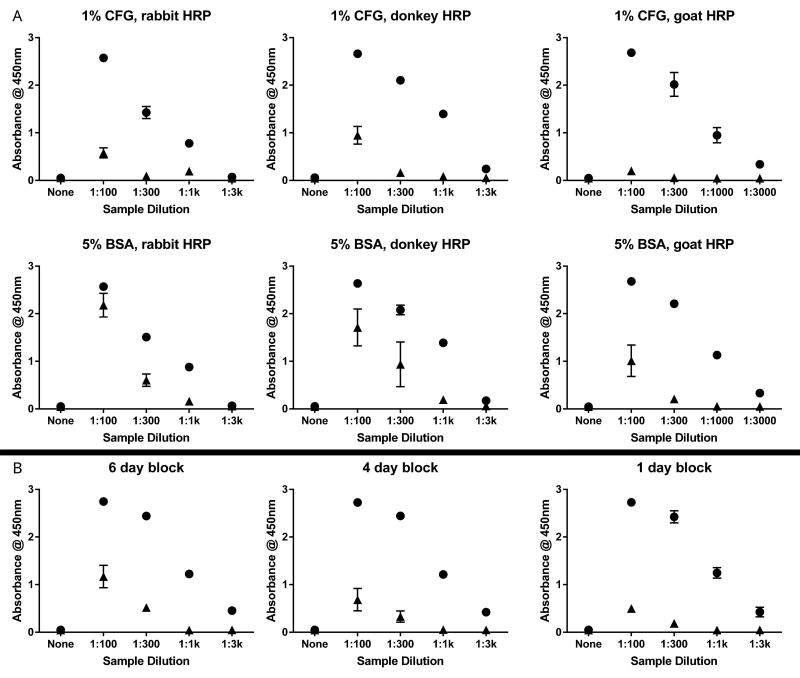
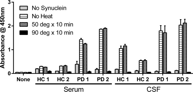
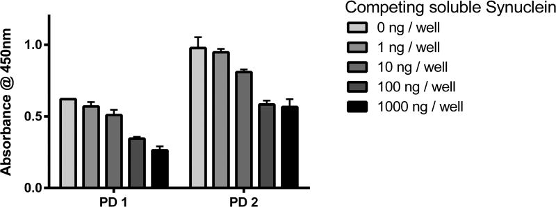
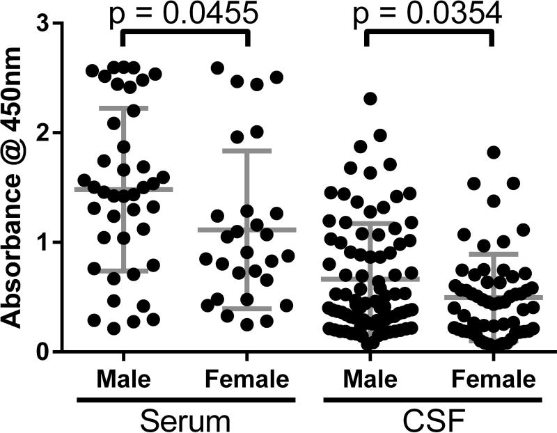
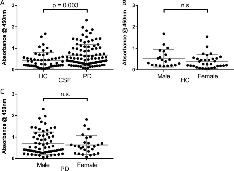
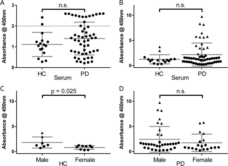
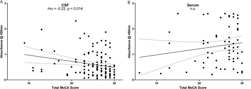
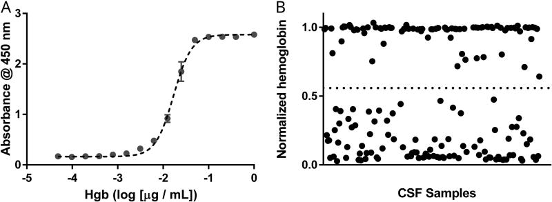
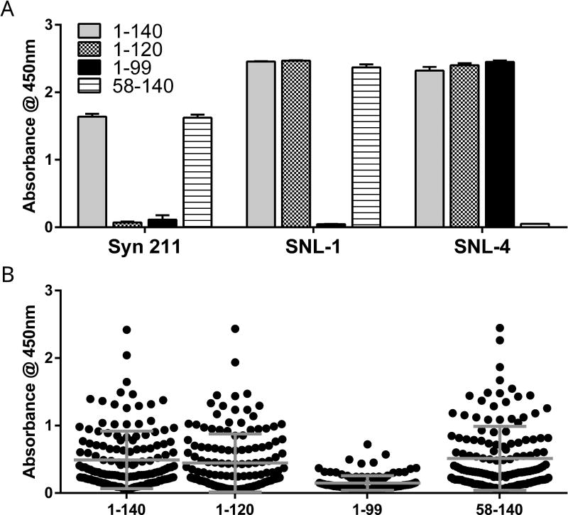
Biomarkers for α-synuclein are needed for diagnosis and prognosis in Parkinson's disease (PD). Endogenous auto-antibodies to α-synuclein could serve as biomarkers for underlying synucleinopathy, but previous assessments of auto-antibodies have shown variability and inconsistent clinical correlations. We hypothesized that auto-antibodies to α-synuclein could be diagnostic for PD and explain its clinical heterogeneity. To test this hypothesis, we developed an enzyme-linked immunosorbent assay for measuring α-synuclein auto-antibodies in human samples. We evaluated 69 serum samples (16 healthy controls (HC) and 53 PD patients) and 145 CSF samples (52 HC and 93 PD patients) from our Institution. Both serum and CSF were available for 24 participants. Males had higher auto-antibody levels than females in both fluids. CSF auto-antibody levels were significantly higher in PD patients as compared with HC, whereas serum levels were not significantly different. CSF auto-antibody levels did not associate with amyloid-β1-42 , total tau, or phosphorylated tau. CSF auto-antibody levels correlated with performance on the Montreal Cognitive Assessment, even when controlled for CSF amyloidβ1-42 . CSF hemoglobin levels, as a proxy for contamination of CSF by blood during lumbar puncture, did not influence these observations. Using recombinant α-synuclein with N- and C-terminal truncations, we found that CSF auto-antibodies target amino acids 100 through 120 of α-synuclein. We conclude that endogenous CSF auto-antibodies are significantly higher in PD patients as compared with HC, suggesting that they could indicate the presence of underlying synucleinopathy. These auto-antibodies associate with poor cognition, independently of CSF amyloidβ1-42 , and target a select C-terminal region of α-synuclein. Read the Editorial Highlight for this article on page 433.

Keywords: Parkinson's disease; auto-antibody; biomarker; neurodegeneration; α-synuclein.


Conflict of interest statement

Conflicts of Interest

The authors declare that they have no competing interests.

Figures

Figure 1
Figure 1. Optimization of ELISA for α-syn AAb detection
Dilute serum was applied to 384-well ELISA plates either coated with recombinant α-syn or not coated. (A) Plates were blocked with either 1% cold fish gelatin (top row) or 5% bovine serum albumin (middle row) overnight at 4 °C. Binding was detected using anti-human IgG HRP-conjugates raised in rabbit (left column), donkey (middle column), or goat (right column). (B) Plates were blocked with 1% cold fish gelatin at 4°C for either 6 days (left), 4 days (middle), or 1 day (right). Additional blocking solutions (1% bovine serum albumin, 5% milk in PBS, several commercial blocking solutions) did not demonstrate any improvement over 1% cold fish gelatin. Each data point represents three technical replicates (n=3 wells). Wells coated with α-syn denoted by closed circles. Uncoated wells denoted by closed triangles. BSA = bovine serum albumin; CFG = cold fish gelatin; HRP = horseradish peroxidase conjugate.
Figure 2
Figure 2. Heat inactivates immunoglobulins detected in α-syn AAb ELISA
Diluted serum or CSF of two HC and two PD patients were prepared in PBS and heated to 50°C, 90°C, or kept on ice for 10 minutes prior to addition to ELISA. Activity on ice was similar to activity following incubation at 50°C for 10 minutes. In contrast, incubation at 90°C for 10 minutes denatured the immunoglobulins in the sample and ELISA activity was lost. Each column represents mean ± standard deviation of three technical replicates (n=3 wells). HC = healthy control; PD = Parkinson’s disease.
Figure 3
Figure 3. Pre-incubation with soluble α-syn reveals low-affinity AAb interactions
Diluted serum of two PD patients were prepared in PBS along with increasing concentrations of soluble α-syn monomer. This preparation was incubated at room temperature for two hours on an orbital shaker before analysis in ELISA. Activity was reduced to 92% (PD1) and 97% (PD2) with 1 ng soluble α-syn monomer, 82% and 83% with 10 ng, 56% and 60% with 100 ng, and 43% and 58% with 1000 ng α-syn in the pre-incubation step. Each column represents mean ± standard deviation of three technical replicates (n=3 wells). PD = Parkinson’s disease.
Figure 4
Figure 4. Auto-antibodies to α-syn are higher in males than females
Males had significantly higher CSF levels of α-syn AAbs (0.664 ± 0.51) than females (0.497 ± 0.40) (t143 = 2.12, p = 0.035). Similarly, levels of AAbs were higher in serum in males (1.481 ± 0.74) than in females (1.114 ± 0.72) (t67 = 2.04, p = 0.046). CSF samples were diluted 1:5 and serum samples were diluted 1:500. Each data point depicts mean ± standard deviation of three samples per assay, each assay having three technical replicates (n=9 wells). Horizontal lines indicate group mean and standard deviations.
Figure 5
Figure 5. CSF auto-antibodies to α-syn are higher in Parkinson’s disease patients as compared to healthy controls
(A) Levels of α-syn AAbs were significantly higher in CSF from PD patients (0.681 ± 0.50) as compared to CSF from HC (0.444 ± 0.36) (t143 = 2.98, p = 0.003). (B) Male HC had higher levels (0.537 ± 0.41) as compared to female HC (0.385 ± 0.33), which was not statistically significant (p = 0.14). (B) Male PD patients also had higher levels (0.702 ± 0.53) as compared to female PD patients (0.629 ± 0.44), which was not statistically significant (p = 0.53). Each data point depicts mean ± standard deviation of three samples per assay, each assay having three technical replicates (n=9 wells). Horizontal lines indicate mean and standard deviations.
Figure 6
Figure 6. Serum auto-antibodies to α-syn are similar between Parkinson’s disease patients and healthy controls
(A) In serum samples diluted 1:500, α-syn AAb levels were not significantly different between HC (1.114 ± 0.58) and PD patients (1.398 ± 0.79) (p = 0.19). (B) Sixteen serum samples re-analyzed at 1:2000 dilution and AAb levels were imputed (closed triangles). Although PD α-syn AAb levels were higher (2.190 ± 2.30) than HC levels (1.250 ± 0.91) when substituting imputed values, this difference was still not statistically significant (p = 0.12). (C) Male HC had significantly higher imputed levels (1.805 ± 1.11) as compared to female HC (0.818 ± 0.37) (t14 = 2.51, p = 0.025). (D) Male PD patients had higher levels (2.440 ± 2.55) than female PD patients (1.744 ± 1.73) that was not significantly different (p = 0.295). Each data point depicts mean ± standard deviation of three samples per assay, each assay having three technical replicates (n=9 wells). Horizontal lines indicate mean and standard deviations. Closed triangles indicate imputed values.
Figure 7
Figure 7. Higher levels of α-syn auto-antibodies in CSF correlate with lower global cognitive performance
Higher levels of CSF α-syn AAbs significantly correlated with lower total score on the MoCA (r = −0.22, p = 0.014), which remained significant when controlled for age and education (see text). In contrast, there was no significant correlation between serum α-syn AAb level and total MoCA score (p = 0.21). Each data point depicts mean of three samples per assay, each assay having three technical replicates (n=9 wells). Each serum data point depicts un-adjusted values. Regression line and 95% confidence band shown.
Figure 8
Figure 8. Hemoglobin measures in CSF using ELISA
CSF diluted 1:5 and a pooled serum sample diluted 1:5000 for normalization were analyzed using a commercially available ELISA for hemoglobin. (A) Representative standard curve with a hemoglobin calibrator fit with four-parameter logistic dose-response curve (Sy.x. = 0.072). (B) Given a narrow dynamic range of this assay, an arbitrary hemoglobin level of 0.558 absorbance units (horizontal dotted line) was selected to dichotomize samples as hemoglobin-absent vs hemoglobin-present. Each data point represents three technical replicates (n=3 wells). Hgb = hemoglobin.
Figure 9
Figure 9. Recruitment of CSF α-syn auto-antibodies is reduced by select C-terminal truncation of α-syn
Full-length or truncated α-syn were adsorbed for ELISA. (A) Mouse monoclonal antibody Syn 211 or rabbit polyclonal antibodies SNL-1 and SNL-4 did not detect α-syn in which truncations removed the binding epitope of each antibody. There was no significant difference in absorbance between the two preparations (full length and a.a 58–140) detected by Syn 211 (p = 0.69). SNL-1 detected slightly less of the N-terminal truncated α-syn than either the full-length or the a.a. 1–120 preparation (p = 0.016 and p = 0.008) (overall effect F2,6 = 12.84, p = 0.007 by one-way ANOVA when excluding a.a. 1–99). SNL-4 detected slightly more of the a.a. 1–99 preparation than full-length (p = 0.013) but there was otherwise no difference between full-length and a.a. 1–120 or between a.a. 1–120 and a.a. 1–99 (overall effect F2,6 = 9.23, p = 0.015 by one-way ANOVA when excluding a.a. 58–140). Each column represents mean ± standard deviation of three technical replicates (n=3 wells). (B) CSF α-syn AAb levels were significantly lower when C-terminal truncated (a.a. 1–99) α-syn was used in ELISA as compared to full-length α-syn (p < 0.001), partial C-terminal truncated α-syn (a.a. 1–120, p < 0.001), or N-terminal truncated α-syn (a.a. 58–140, p < 0.001), (overall effect F3,564 = 27.32, p < 0.0001 by one-way ANOVA). There was no significant difference in average CSF α-syn AAb levels detected by full-length vs a.a 1–120 (p = 0.75), full-length vs a.a. 58–140 (p = 0.96), or a.a 1–120 vs 58–140 (p = 0.44). Each data point depicts mean of three samples per assay, each assay having three technical replicates (n=9 wells). Horizontal lines depict mean and standard deviations

Comment in

References

    1. Adler CH, Connor DJ, Hentz JG, Sabbagh MN, Caviness JN, Shill HA, Noble B, Beach TG. Incidental Lewy body disease: clinical comparison to a control cohort. Movement disorders : official journal of the Movement Disorder Society. 2010;25:642–646. - PMC - PubMed
    1. Akhtar RS, Stern MB. New concepts in the early and preclinical detection of Parkinson's disease: therapeutic implications. Expert review of neurotherapeutics. 2012;12:1429–1438. - PubMed
    1. Alves G, Lange J, Blennow K, Zetterberg H, Andreasson U, Forland MG, Tysnes OB, Larsen JP, Pedersen KF. CSF Abeta42 predicts early-onset dementia in Parkinson disease. Neurology. 2014;82:1784–1790. - PubMed
    1. Beach TG, Adler CH, Dugger BN, et al. Submandibular gland biopsy for the diagnosis of Parkinson disease. Journal of neuropathology and experimental neurology. 2013;72:130–136. - PMC - PubMed
    1. Beach TG, Adler CH, Sue LI, et al. Multi-organ distribution of phosphorylated alpha-synuclein histopathology in subjects with Lewy body disorders. Acta neuropathologica. 2010;119:689–702. - PMC - PubMed

Publication types

MeSH terms