Review
. 2018 Apr;22(4):350-364.
doi: 10.1016/j.tics.2018.01.010. Epub 2018 Feb 28.

How to Characterize the Function of a Brain Region

Affiliations
Review

How to Characterize the Function of a Brain Region

Sarah Genon et al. Trends Cogn Sci. 2018 Apr.

Abstract

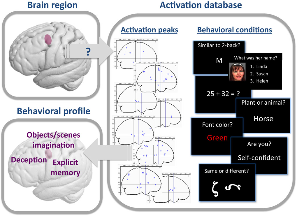
Many brain regions have been defined, but a comprehensive formalization of each region's function in relation to human behavior is still lacking. Current knowledge comes from various fields, which have diverse conceptions of 'functions'. We briefly review these fields and outline how the heterogeneity of associations could be harnessed to disclose the computational function of any region. Aggregating activation data from neuroimaging studies allows us to characterize the functional engagement of a region across a range of experimental conditions. Furthermore, large-sample data can disclose covariation between brain region features and ecological behavioral phenotyping. Combining these two approaches opens a new perspective to determine the behavioral associations of a brain region, and hence its function and broader role within large-scale functional networks.

Keywords: BrainMap; Functional specialization; MRI; Neurosynth; brain mapping; data-driven.


Figures

Figure 1.
Figure 1.. Illustration of Behavioral Functional Profiling for the Left Rostral Dorsal Premotor Cortex (PMd).
Activation databases (such as BrainMap and Neurosynth) contain a collection of activation peaks that have been reported in stereotactic space in scientific papers, as well as information on behavioral conditions associated with these peaks (based on the behavioral task that the participants had to perform in the MRI scanner). For a given brain region-of-interest (here the rostral left PMd), we searched among all the peaks of activation reported in the BrainMap database for those that were located in this region. In this database, the behavioral condition related to each peak is specified in terms of behavioral paradigms and behavior domains. Examining the behavioral paradigms and behavioral domains in which the peaks of activation were consistently reported in the region-of-interest allowed us to establish a behavioral profile of this region. As illustrated in the left inferior panel, the left rostral PMd was found to be activated in experimental tasks probing explicit memory, object or scene imagination, and deception [78]. The face used to illustrate a recognition paradigm comes from the Glasgow Unfamiliar Face Database (GUFD) [104].
Figure 2.
Figure 2.. Structural Brain–Behavior Correlation Approach.
(A) Manhattan plot relating a set of cerebral measures derived from anatomical images to non-brain phenotypical variables (1100 variables clustered into major groups along the x-axis) in the UK BioBank cohort. For each variable, the significance of the cross-subject correlation with each brain measure set is plotted vertically in units of −log 10 [86]. (B) Positive correlation between conscientiousness scores and gray-matter volume (GMV) in the cuneus in males only, within a sample from the Human Connectome Project (HCP) [65]. (C) Significant relationship between hippocampal volume and immediate recall performance at the Rey Auditory Verbal Learning Test interacting with gender in a sample of participants with mild cognitive impairment (MCI) from the Alzheimer’s Disease Neuroimaging Initiative (ADNI) [87]. Abbreviations: Bonf, Bonferroni correction; FDR, false discovery rate; n, number of participants.

References

    1. Amunts K et al. (2014) Interoperable atlases of the human brain. Neuroimage 99, 525–532 - PubMed
    1. Lorenz S et al. (2017) Two new cytoarchitectonic areas on the human mid-fusiform gyrus. Cereb. Cortex 27, 373–385 - PMC - PubMed
    1. Toga AW et al. (2006) Towards multimodal atlases of the human brain. Nat. Rev. Neurosci 7, 952–966 - PMC - PubMed
    1. Eickhoff SB et al. (2007) Assignment of functional activations to probabilistic cytoarchitectonic areas revisited. Neuroimage 36, 511–521 - PubMed
    1. Yeo BT et al. (2011) The organization of the human cerebral cortex estimated by intrinsic functional connectivity. J. Neurophysiol 106, 1125–1165 - PMC - PubMed

Publication types