. 2018 Aug;138(8):1862-1870.
doi: 10.1016/j.jid.2018.02.023. Epub 2018 Mar 6.

Alteration of Extracellular Nucleotide Metabolism in Pseudoxanthoma Elasticum

Affiliations

Alteration of Extracellular Nucleotide Metabolism in Pseudoxanthoma Elasticum

Gilles Kauffenstein et al. J Invest Dermatol. 2018 Aug.

Abstract

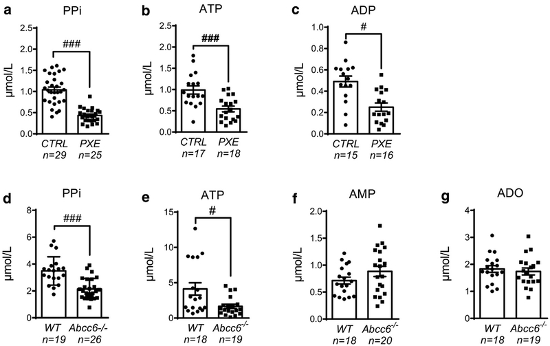
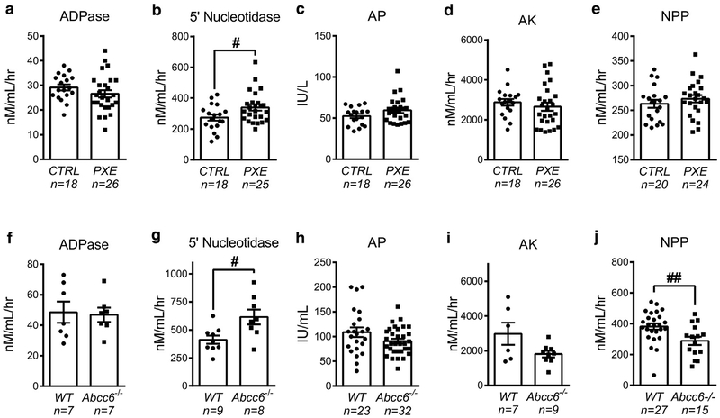
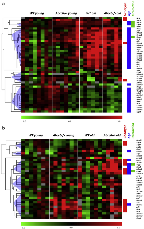
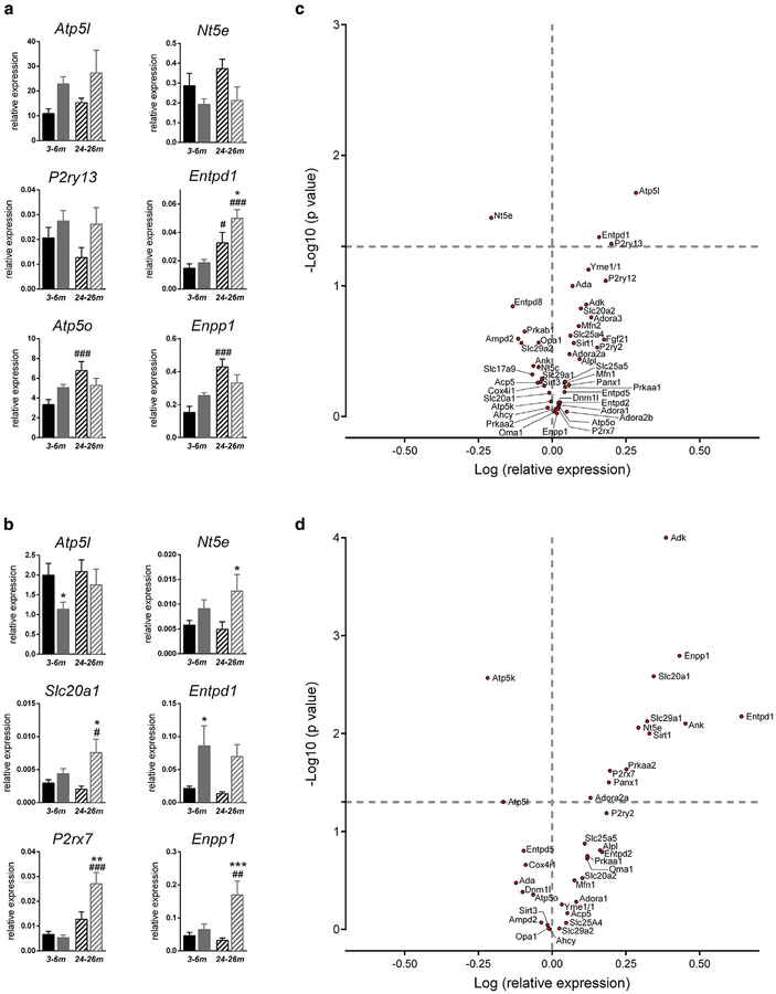
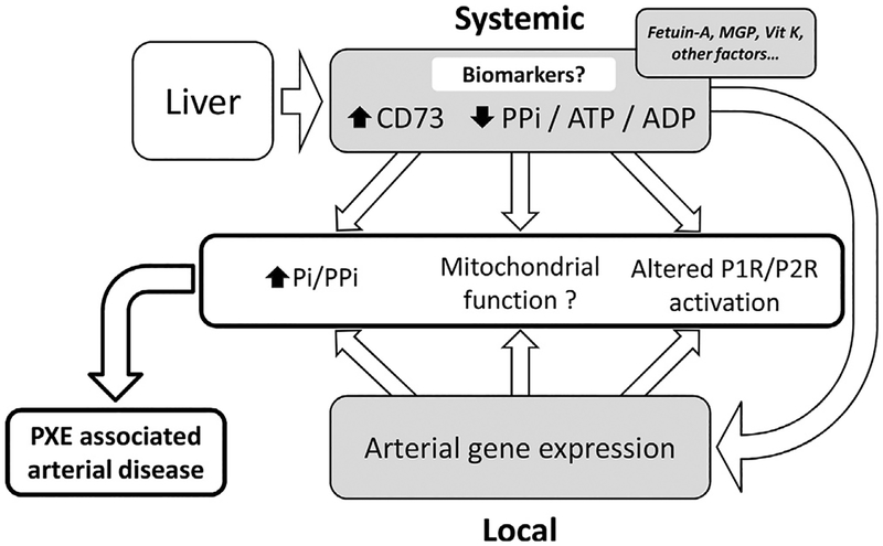
Pseudoxanthoma elasticum (PXE) is a rare genetic condition primarily caused by hepatic ABCC6 transporter dysfunction. Most clinical manifestations of PXE are due to premature calcification of elastic fibers. However, the vascular impact of PXE is pleiotropic and remains ill defined. ABCC6 expression has recently been associated with cellular nucleotide export. We studied the impact of ABCC6 deficiency on blood levels of adenosine triphosphate and related metabolites and on soluble nucleotidase activities in PXE patients and Abcc6-/- mice. In addition, we investigated the expression of genes encoding ectocellular purinergic signaling proteins in mouse liver and aorta. Plasma adenosine triphosphate and pyrophosphate levels were significantly reduced in PXE patients and in Abcc6-/- mice, whereas adenosine concentration was not modified. Moreover, 5'-nucleotidase/CD73 activity was increased in the serum of PXE patients and Abcc6-/- mice. Consistent with alterations of purinergic signaling, the expression of genes involved in purine and phosphate transport/metabolism was dramatically modified in Abcc6-/- mouse aorta, with much less impact on the liver. ABCC6 deficiency causes impaired vascular homeostasis and tissue perfusion. Our findings suggest that these alterations are linked to changes in extracellular nucleotide metabolism that are remote from the liver. This opens new perspectives for the understanding of PXE pathophysiology.


Conflict of interest statement

CONFLICT OF INTEREST

The authors state no conflict of interest.

Figures

Figure 1.
Figure 1.. Quantification of plasma adenine nucleotide, ADO, and PPi levels during ABCC6 deficiency.
Concentrations of (a) PPi, (b) ATP, and (c) ADP were determined in blood plasma from healthy control (CTRL) and PXE individuals. (d) PlasmaPPi, (e) ATP, (f) AMP, and (g) adenosine were measured in WT and Abcc6−/− mice. Data represent the mean ± standard error of the mean of the indicated number of plasma samples. #P < 0.05, ###P < 0.005. ADO, adenosine; AMP, adenosine monophosphate; ATP, adenosine triphosphate; CTRL, control; PPi, pyrophosphate; PXE, pseudoxanthoma elasticum; WT, wild type.
Figure 2.
Figure 2.. Changes in soluble nucleotide-converting activities in PXE patients and Abcc6−/− mice.
The activities of key soluble enzymes, (a, f) NTPDase1/CD39 (ADPase), (b, g) ecto-5′-nucleotidase/CD73, (c, h) alkaline phosphatase (AP), (d, i) adenylate kinase (AK), and (e, j) nucleotide pyrophosphatase phosphodiesterase (NPP) were determined in (a–e) the serum of healthy control individuals and PXE patients and in (f–j) Abcc6−/− and WT mice. Data represent the mean ± standard error of the mean of the indicated number of serum samples. #P < 0.05; ##P < 0.01. CTRL, control; IU, international unit; nM/mL/hr, nano molar/milliliter/hour; PXE, pseudoxanthoma elasticum.
Figure 3.
Figure 3.. Gene expression pattern in liver and aorta of Abcc6−/− mice.
Heat map of gene expression in the (a) liver and (b) aorta of young (5 months, n = 7) and old (24 months, n = 6, except for Abcc6−/−aorta, for which n = 5) Abcc6+/+ and Abcc6−/− mice. Results represent color-coded expression relative to the control group (WT young). On the right are noted genes that are differently expressed (P < 0.05, two-way analysis of variance) according to genotype (red box) and to age (blue box) and the significance of the interaction between the two factors (green box). WT, wild type.
Figure 4.
Figure 4.. Gene expression pattern in liver and aorta of Abcc6−/− mice.
Bar graphs illustrating representative gene expression pattern in young andold WT and Abcc6−/− mice in the (a) liver and (b) aorta. Genes represented were selected according to (i) the significant effect of Abcc6 genotype and(ii) the significance of aging/genotype interaction. Black bars indicate WT mice, and gray bars indicate Abcc6−/− mice; solid fill indicatesadult mice, and hatched fill indicates old mice. Significant differences according to age (#) or genotype (*) (P < 0.05). Volcano plots showing the distribution of relative gene expression (log of fold changes) and P-values (log10) in (c) Abcc6−/− mice liver (n = 13) and (d) aorta (n = 13 WT and n = 12 Abcc6−/−). Data are the mean of five to seven values obtained from different animals. Values of log P above 1.3 (P < 0.05) are statistically significant. #P < 0.05; ###P < 0.001; *P < 0.05; **P < 0.01; ***P < 0.001. WT, wild type.
Figure 5.
Figure 5.. Proposal of a summary diagram for the pathophysiology of PXE.
In the context of liver (central) ABCC6 deficiency, plasma levels of PPi and adenine nucleotides (ATP, ADP) are decreased and 5′-nucleotidase activity is increased each of these, representing potential disease biomarkers. ABCC6 deficiency is also associated with modifications of purine and phosphate gene expression in (remote) affected tissues like arteries. Both systemic and local alterations in purine and phosphate metabolism contribute to PXE-associated peripheral arterial disease. ADP, adenosine diphosphate; ATP, adenosine triphosphate; MGP, matrix gla protein; Pi, inorganic phosphate; PPi, pyrophosphate; PXE, pseudoxanthoma elasticum; Vit, vitamin.

Similar articles

Cited by

  • Multi-ancestry genome-wide study identifies effector genes and druggable pathways for coronary artery calcification.
    Kavousi M, Bos MM, Barnes HJ, Lino Cardenas CL, Wong D, Lu H, Hodonsky CJ, Landsmeer LPL, Turner AW, Kho M, Hasbani NR, de Vries PS, Bowden DW, Chopade S, Deelen J, Benavente ED, Guo X, Hofer E, Hwang SJ, Lutz SM, Lyytikäinen LP, Slenders L, Smith AV, Stanislawski MA, van Setten J, Wong Q, Yanek LR, Becker DM, Beekman M, Budoff MJ, Feitosa MF, Finan C, Hilliard AT, Kardia SLR, Kovacic JC, Kral BG, Langefeld CD, Launer LJ, Malik S, Hoesein FAAM, Mokry M, Schmidt R, Smith JA, Taylor KD, Terry JG, van der Grond J, van Meurs J, Vliegenthart R, Xu J, Young KA, Zilhão NR, Zweiker R, Assimes TL, Becker LC, Bos D, Carr JJ, Cupples LA, de Kleijn DPV, de Winther M, den Ruijter HM, Fornage M, Freedman BI, Gudnason V, Hingorani AD, Hokanson JE, Ikram MA, Išgum I, Jacobs DR Jr, Kähönen M, Lange LA, Lehtimäki T, Pasterkamp G, Raitakari OT, Schmidt H, Slagboom PE, Uitterlinden AG, Vernooij MW, Bis JC, Franceschini N, Psaty BM, Post WS, Rotter JI, Björkegren JLM, O'Donnell CJ, Bielak LF, Peyser PA, Malhotra R, van der Laan SW, Miller CL. Kavousi M, et al. Nat Genet. 2023 Oct;55(10):1651-1664. doi: 10.1038/s41588-023-01518-4. Epub 2023 Sep 28. Nat Genet. 2023. PMID: 37770635 Free PMC article.
  • Plasma inorganic pyrophosphate and alkaline phosphatase in patients with pseudoxanthoma elasticum.
    Sánchez-Tévar AM, García-Fernández M, Murcia-Casas B, Rioja-Villodres J, Carrillo JL, Camacho M, Van Gils M, Sánchez-Chaparro MA, Vanakker O, Valdivielso P. Sánchez-Tévar AM, et al. Ann Transl Med. 2019 Dec;7(24):798. doi: 10.21037/atm.2019.12.73. Ann Transl Med. 2019. PMID: 32042814 Free PMC article.
  • Histological Findings in the Eyes of Abcc6 Knockout Rat Model of Pseudoxanthoma Elasticum.
    Sehgal A, Milman T, Li Q, Pulido JS. Sehgal A, et al. Transl Vis Sci Technol. 2024 Apr 2;13(4):29. doi: 10.1167/tvst.13.4.29. Transl Vis Sci Technol. 2024. PMID: 38656313 Free PMC article.
  • A Plasma Pyrophosphate Cutoff Value for Diagnosing Pseudoxanthoma Elasticum.
    Rubera I, Clotaire L, Laurain A, Destere A, Martin L, Duranton C, Leftheriotis G. Rubera I, et al. Int J Mol Sci. 2024 Jun 13;25(12):6502. doi: 10.3390/ijms25126502. Int J Mol Sci. 2024. PMID: 38928212 Free PMC article.
  • Metabolites and metabolism in vascular calcification: links between adenosine signaling and the methionine cycle.
    Behzadi P, St Hilaire C. Behzadi P, et al. Am J Physiol Heart Circ Physiol. 2024 Dec 1;327(6):H1361-H1375. doi: 10.1152/ajpheart.00267.2024. Epub 2024 Oct 25. Am J Physiol Heart Circ Physiol. 2024. PMID: 39453431 Free PMC article. Review.

References

    1. Aessopos A, Savvides P, Stamatelos G, Rombos I, Tassiopoulos T, Karagiorga M, et al. Pseudoxanthoma elasticum-like skin lesions and angioid streaks in beta-thalassemia. Am J Hematol 1992;41:159e64. - PubMed
    1. Beck K, Hayashi K, Nishiguchi B, Le Saux O, Hayashi M, Boyd CD. The distribution of Abcc6 in normal mouse tissues suggests multiple functions for this ABC transporter. J Histochem Cytochem 2003;51:887e902. - PubMed
    1. Bergen AA, Plomp AS, Schuurman EJ, Terry S, Breuning M, Dauwerse H, et al. Mutations in ABCC6 cause Pseudoxanthoma elasticum. Nat Genet 2000;25:228e31. - PubMed
    1. Boraldi F, Annovi G, Bartolomeo A, Quaglino D. Fibroblasts from patients affected by Pseudoxanthoma elasticum exhibit an altered PPi metabolism and are more responsive to pro-calcifying stimuli. J Dermatol Sci 2014a;74: 72e80. - PubMed
    1. Boraldi F, Bartolomeo A, Li Q, Uitto J, Quaglino D. Changes in dermal fibroblasts from Abcc6(−/−) mice are present before and after the onset of ectopic tissue mineralization. J Invest Dermatol 2014b;134:1855e61. - PMC - PubMed

Publication types

MeSH terms

LinkOut - more resources