

New genetic tools for the in vivo study of hematopoietic stem cell function
- PMID: 29501466
- PMCID: PMC5899657
- DOI: 10.1016/j.exphem.2018.02.004
New genetic tools for the in vivo study of hematopoietic stem cell function
Abstract
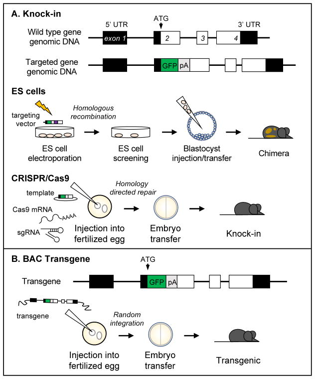
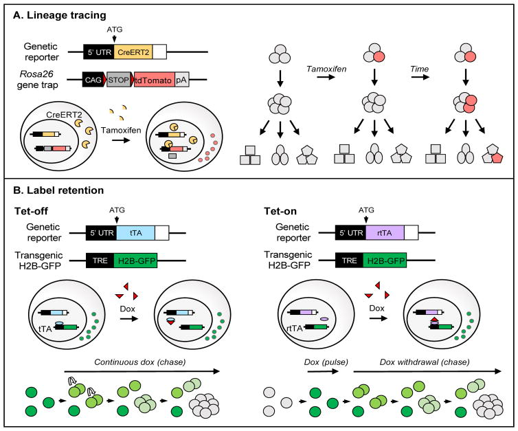
The production of blood cells is dependent on the activity of a rare stem cell population that normally resides in the bone marrow (BM) of the organism. These hematopoietic stem cells (HSCs) have the ability to both self-renew and differentiate, ensuring this lifelong hematopoiesis. Determining the regulation of HSC functions should thus provide critical insight to advancing regenerative medicine. Until quite recently, HSCs were primarily studied using in vitro studies and transplantations into immunodeficient hosts. Indeed, the definition of a bona fide HSC is its ability to reconstitute lymphopenic hosts. In this review, we discuss the development of novel, HSC-specific genetic reporter systems that enable the prospective identification of HSCs and the study of their functions in the absence of transplantation. Coupled with additional technological advances, these studies are now defining the fundamental properties of HSCs in vivo. Furthermore, complex cellular and molecular mechanisms that regulate HSC dormancy, self-renewal, and differentiation are being identified and further dissected. These novel reporter systems represent a major technological advance for the stem cell field and allow new questions to be addressed.
Copyright © 2018 ISEH – Society for Hematology and Stem Cells. Published by Elsevier Inc. All rights reserved.
Figures
Similar articles
-
Hematopoietic Stem Cells, Their Niche, and the Concept of Co-Culture Systems: A Critical Review.J Stem Cells. 2015;10(1):13-31. J Stem Cells. 2015. PMID: 26665935 Review.
-
Mechanisms controlling hematopoietic stem cell functions during normal hematopoiesis and hematological malignancies.Wiley Interdiscip Rev Syst Biol Med. 2011 Nov-Dec;3(6):681-701. doi: 10.1002/wsbm.145. Epub 2011 Mar 15. Wiley Interdiscip Rev Syst Biol Med. 2011. PMID: 21412991 Review.
-
Quiescent human hematopoietic stem cells in the bone marrow niches organize the hierarchical structure of hematopoiesis.Stem Cells. 2008 Dec;26(12):3228-36. doi: 10.1634/stemcells.2008-0552. Epub 2008 Sep 11. Stem Cells. 2008. PMID: 18787204
-
Niche heterogeneity in the bone marrow.Ann N Y Acad Sci. 2016 Apr;1370(1):82-96. doi: 10.1111/nyas.13016. Epub 2016 Mar 25. Ann N Y Acad Sci. 2016. PMID: 27015419 Free PMC article. Review.
-
Differentiation-based model of hematopoietic stem cell functions and lineage pathways.Blood. 2018 Sep 13;132(11):1106-1113. doi: 10.1182/blood-2018-03-791517. Epub 2018 Jul 24. Blood. 2018. PMID: 30042097 Free PMC article. Review.
Cited by
-
Lin- PU.1dim GATA-1- defines haematopoietic stem cells with long-term multilineage reconstitution activity.Cell Prolif. 2023 Nov;56(11):e13490. doi: 10.1111/cpr.13490. Epub 2023 May 5. Cell Prolif. 2023. PMID: 37147872 Free PMC article.
-
Do haematopoietic stem cells age?Nat Rev Immunol. 2020 Mar;20(3):196-202. doi: 10.1038/s41577-019-0236-2. Epub 2019 Nov 18. Nat Rev Immunol. 2020. PMID: 31740804 Free PMC article. Review.
-
New insights into hematopoietic differentiation landscapes from single-cell RNA sequencing.Blood. 2019 Mar 28;133(13):1415-1426. doi: 10.1182/blood-2018-08-835355. Epub 2019 Feb 6. Blood. 2019. PMID: 30728144 Free PMC article. Review.
-
Hematopoietic stem cell fate through metabolic control.Exp Hematol. 2018 Aug;64:1-11. doi: 10.1016/j.exphem.2018.05.005. Epub 2018 May 25. Exp Hematol. 2018. PMID: 29807063 Free PMC article. Review.
-
Tracking hematopoietic stem cells and their progeny using whole-genome sequencing.Exp Hematol. 2020 Mar;83:12-24. doi: 10.1016/j.exphem.2020.01.004. Epub 2020 Jan 30. Exp Hematol. 2020. PMID: 32007478 Free PMC article.
References
-
- Adolfsson J, et al. Identification of Flt3+ lympho-myeloid stem cells lacking erythro-megakaryocytic potential a revised road map for adult blood lineage commitment. Cell. 2005;121(2):295–306. - PubMed
-
- Kiel MJ, et al. SLAM family receptors distinguish hematopoietic stem and progenitor cells and reveal endothelial niches for stem cells. Cell. 2005;121(7):1109–21. - PubMed
-
- Osawa M, et al. Long-term lymphohematopoietic reconstitution by a single CD34-low/negative hematopoietic stem cell. Science. 1996;273(5272):242–5. - PubMed
Publication types
MeSH terms
Grants and funding
LinkOut - more resources
Full Text Sources
Other Literature Sources
Medical
