

Adipose tissue extrinsic factor: Obesity-induced inflammation and the role of the visceral lymph node
- PMID: 29501838
- PMCID: PMC6461448
- DOI: 10.1016/j.physbeh.2018.02.044
Adipose tissue extrinsic factor: Obesity-induced inflammation and the role of the visceral lymph node
Abstract
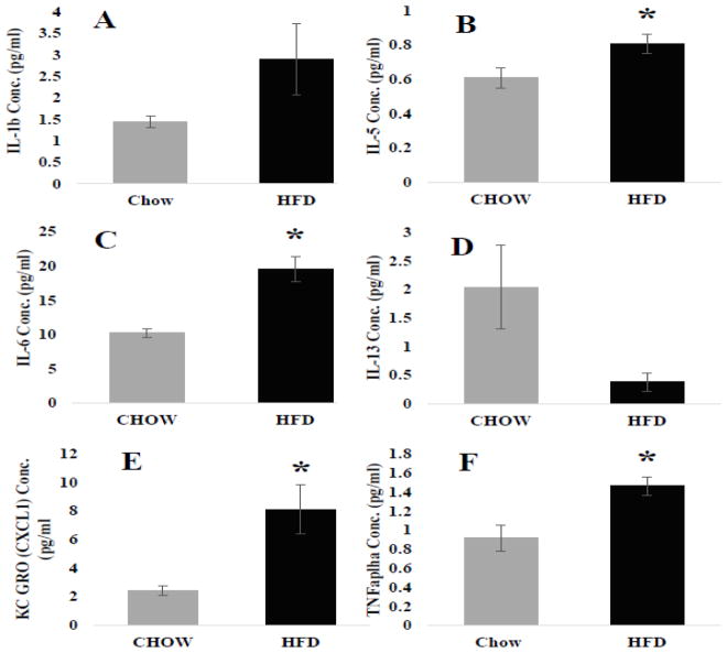
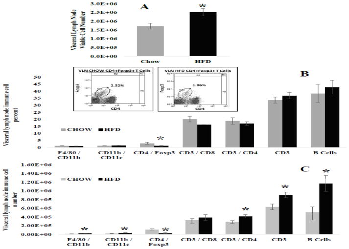
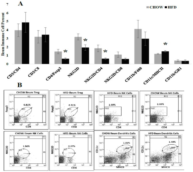
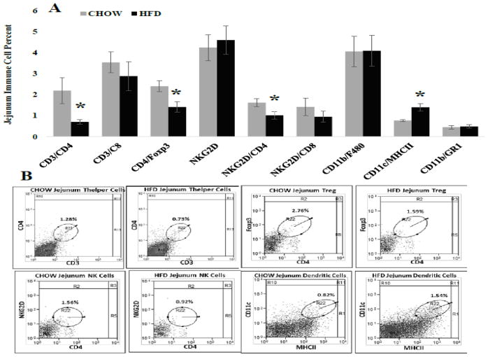
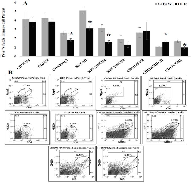
Obesity-related adverse health consequences occur predominately in individuals with upper body fat distribution commonly associated with increased central adiposity. Visceral adipose tissue accumulation is described to be the greatest driver of obesity-induced inflammation, however evidence also supports that the intestines fundamentally contribute to the development of obesity-induced metabolic disease. The visceral adipose depot shares the same vasculature and lymph drainage as the small intestine. We hypothesize that the visceral lymph node, which drains adipose tissue and the gastrointestinal tract, is central to the exacerbation of systemic pro-inflammation. Male C57BL/6 mice were fed CHOW or high fat diet (HFD) for 7 weeks. At termination the mesenteric depot, visceral lymph node and ileum, jejunum and Peyer's patches were collected. Cytokine concentration was determined in adipose tissue whereas immune cell populations where investigated in the visceral lymph node and intestinal segments by flow cytometry. Visceral adipose tissue and the gastrointestinal tract mutually influence immune cells enclosed within the visceral lymph node. HFD increased visceral lymph node immune cell number. This likely resulted from 1.) an increase in immune cells migration from the small intestines likely from activated dendritic cells that travel to the lymph node and 2.) cytokine effluent from visceral adipose tissue that promoted expansion, survival and retention of pro-inflammatory immune cells. Overall, the visceral lymph node, the immune nexus of visceral adipose tissue and the small intestines, likely plays a fundamental role in exacerbation of systemic pro-inflammation by HFD-induced obesity. The research of Tim Bartness greatly enhanced the understanding of adipose tissue regulation. Studies from his laboratory significantly contributed to our awareness of extrinsic factors that influence body fatness levels. Specifically, the work he produced eloquently demonstrated that adipose tissue was more complex than an insulating storage center; it was connected to our brains via the sympathetic and sensory nervous system. Mapping studies demonstrated that adipose tissue both receives and sends information to the brain. Further, his lab demonstrated that nervous system connections contributed to lipolysis, thermogenesis and adipocyte proliferation and growth. The work of Tim Bartness will continue to influence adipose tissue research. As such, Tim Bartness directly inspired the following research. Adipose tissue extrinsic factors are not limited to the peripheral nervous system. The lymphatic system is an additional extrinsic factor that cross talks with adipose tissue, however its role in this context is under emphasized. Here we begin to elucidate how the lymphatic system may contribute to the comorbidities associated with visceral adipose tissue accumulation.
Keywords: Central obesity; Lymphatics; Metabolic disease; Pro-inflammation; Visceral adiposity.
Copyright © 2018 Elsevier Inc. All rights reserved.
Conflict of interest statement
The authors declare that they have no conflicts of interest.
Figures
References
-
- Lamon-Fava S, Wilson PW, Schaefer EJ. Impact of body mass index on coronary heart disease risk factors in men and women. The Framingham Offspring Study. Arterioscler Thromb Vasc Biol. 1996;16(12):1509–15. - PubMed
-
- Miyake T, et al. Body mass index is the most useful predictive factor for the onset of nonalcoholic fatty liver disease: a community-based retrospective longitudinal cohort study. J Gastroenterol. 2013;48(3):413–22. - PubMed
-
- O’Neill S, O’Driscoll L. Metabolic syndrome: a closer look at the growing epidemic and its associated pathologies. Obes Rev. 2015;16(1):1–12. - PubMed
-
- Bjorntorp P. Metabolic implications of body fat distribution. Diabetes Care. 1991;14(12):1132–43. - PubMed
Publication types
MeSH terms
Substances
Grants and funding
LinkOut - more resources
Full Text Sources
Other Literature Sources
Medical
Research Materials
Miscellaneous
