Negative regulation of lens fiber cell differentiation by RTK antagonists Spry and Spred
- PMID: 29501879
- PMCID: PMC5924633
- DOI: 10.1016/j.exer.2018.02.025
Negative regulation of lens fiber cell differentiation by RTK antagonists Spry and Spred
Abstract
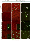
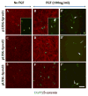
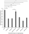
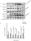
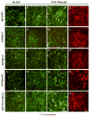
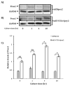
Sprouty (Spry) and Spred proteins have been identified as closely related negative regulators of the receptor tyrosine kinase (RTK)-mediated MAPK pathway, inhibiting cellular proliferation, migration and differentiation in many systems. As the different members of this antagonist family are strongly expressed in the lens epithelium in overlapping patterns, in this study we used lens epithelial explants to examine the impact of these different antagonists on the morphologic and molecular changes associated with fibroblast growth factor (FGF)-induced lens fiber differentiation. Cells in lens epithelial explants were transfected using different approaches to overexpress the different Spry (Spry1, Spry2) and Spred (Spred1, Spred2, Spred3) members, and we compared their ability to undergo FGF-induced fiber differentiation. In cells overexpressing any of the antagonists, the propensity for FGF-induced cell elongation was significantly reduced, indicative of a block to lens fiber differentiation. Of these antagonists, Spry1 and Spred2 appeared to be the most potent among their respective family members, demonstrating the greatest block in FGF-induced fiber differentiation based on the percentage of cells that failed to elongate. Consistent with the reported activity of Spry and Spred, we show that overexpression of Spry2 was able to suppress FGF-induced ERK1/2 phosphorylation in lens cells, as well as the ERK1/2-dependent fiber-specific marker Prox1, but not the accumulation of β-crystallins. Taken together, Spry and Spred proteins that are predominantly expressed in the lens epithelium in situ, appear to have overlapping effects on negatively regulating ERK1/2-signaling associated with FGF-induced lens epithelial cell elongation leading to fiber differentiation. This highlights the important regulatory role for these RTK antagonists in establishing and maintaining the distinct architecture and polarity of the lens.
Keywords: FGF; Lens fiber differentiation; RTK-Antagonists; Spred; Sprouty.
Crown Copyright © 2018. Published by Elsevier Ltd. All rights reserved.
Figures
Similar articles
-
Spred negatively regulates lens growth by modulating epithelial cell proliferation and fiber differentiation.Exp Eye Res. 2019 Jan;178:160-175. doi: 10.1016/j.exer.2018.09.019. Epub 2018 Oct 2. Exp Eye Res. 2019. PMID: 30290165
-
Negative regulation of TGFβ-induced lens epithelial to mesenchymal transition (EMT) by RTK antagonists.Exp Eye Res. 2015 Mar;132:9-16. doi: 10.1016/j.exer.2015.01.001. Epub 2015 Jan 7. Exp Eye Res. 2015. PMID: 25576668
-
Sprouty and Spred temporally regulate ERK1/2-signaling to suppress TGFβ-induced lens EMT.Exp Eye Res. 2022 Jun;219:109070. doi: 10.1016/j.exer.2022.109070. Epub 2022 Apr 9. Exp Eye Res. 2022. PMID: 35413282
-
Fibrosis in the lens. Sprouty regulation of TGFβ-signaling prevents lens EMT leading to cataract.Exp Eye Res. 2016 Jan;142:92-101. doi: 10.1016/j.exer.2015.02.004. Epub 2015 May 21. Exp Eye Res. 2016. PMID: 26003864 Free PMC article. Review.
-
Sprouty proteins, masterminds of receptor tyrosine kinase signaling.Angiogenesis. 2008;11(1):53-62. doi: 10.1007/s10456-008-9089-1. Epub 2008 Jan 25. Angiogenesis. 2008. PMID: 18219583 Review.
Cited by
-
FGF-2 Differentially Regulates Lens Epithelial Cell Behaviour during TGF-β-Induced EMT.Cells. 2023 Mar 7;12(6):827. doi: 10.3390/cells12060827. Cells. 2023. PMID: 36980168 Free PMC article.
-
A possible connection between reactive oxygen species and the unfolded protein response in lens development: From insight to foresight.Front Cell Dev Biol. 2022 Sep 21;10:820949. doi: 10.3389/fcell.2022.820949. eCollection 2022. Front Cell Dev Biol. 2022. PMID: 36211466 Free PMC article. Review.
-
Conditional Ablation of Spred1 and Spred2 in the Eye Lens Negatively Impacts Its Development and Growth.Cells. 2024 Feb 6;13(4):290. doi: 10.3390/cells13040290. Cells. 2024. PMID: 38391903 Free PMC article.
-
Generation of Lens Progenitor Cells and Lentoid Bodies from Pluripotent Stem Cells: Novel Tools for Human Lens Development and Ocular Disease Etiology.Cells. 2022 Nov 6;11(21):3516. doi: 10.3390/cells11213516. Cells. 2022. PMID: 36359912 Free PMC article. Review.
-
Changes in DNA methylation hallmark alterations in chromatin accessibility and gene expression for eye lens differentiation.Epigenetics Chromatin. 2022 Mar 5;15(1):8. doi: 10.1186/s13072-022-00440-z. Epigenetics Chromatin. 2022. PMID: 35246225 Free PMC article.
References
-
- Ball LJ, Jarchau T, Oschkinat H, Walter U. EVH1 domains: structure, function and interactions. FEBS Lett. 2002;513:45–52. - PubMed
-
- Bennett AM, Tonks NK. Regulation of distinct stages of skeletal nuscle differentiation by mitogen-activated protein kinases. Science. 1997;278:1288–1291. - PubMed
-
- Bundschu K, Walter U, Schuh K. Getting a first clue about SPRED functions. Bioessays. 2007;29:897–907. - PubMed
Publication types
MeSH terms
Substances
Grants and funding
LinkOut - more resources
Full Text Sources
Other Literature Sources
Molecular Biology Databases
Research Materials
Miscellaneous

