

Delayed diagnosis of post-surgical pyoderma gangrenosum: A multicenter case series and review of literature
- PMID: 29501933
- PMCID: PMC5910501
- DOI: 10.1016/j.ijscr.2018.02.026
Delayed diagnosis of post-surgical pyoderma gangrenosum: A multicenter case series and review of literature
Abstract
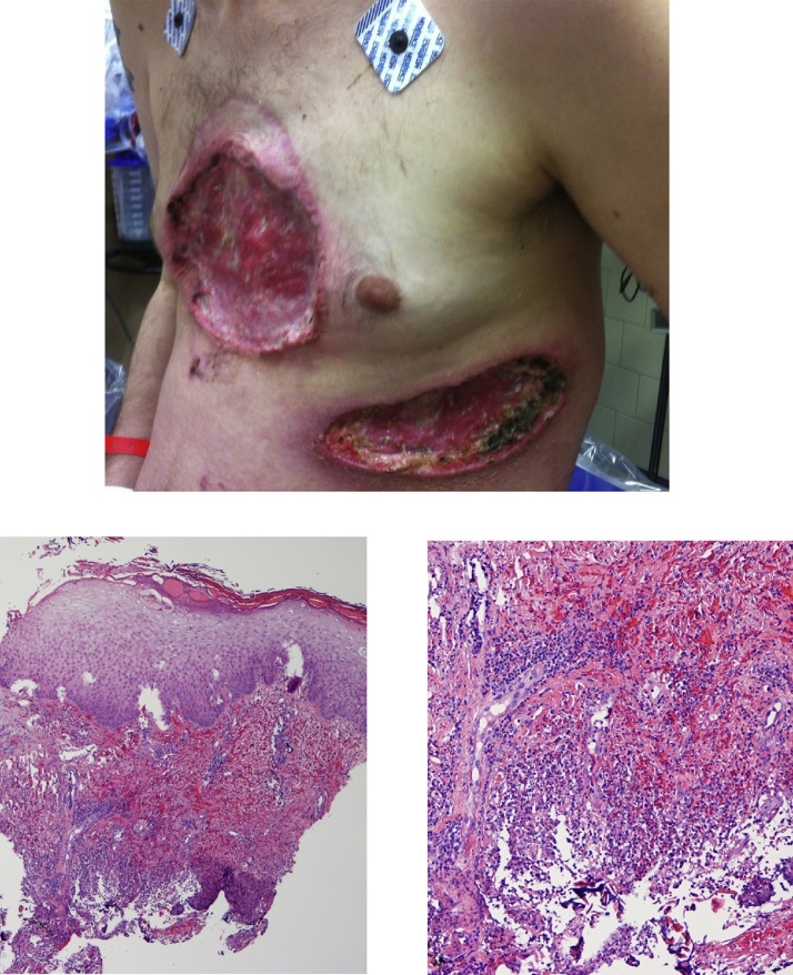
Introduction: Pyoderma gangrenosum is a chronic neutrophilic dermatosis which can occur following trauma or surgery and can mimic infection. Surgical intervention can lead to progression of disease.
Presentation of cases: This case series describes 3 cases of post-surgical pyoderma gangrenosum with delayed diagnosis from two large medical centers.
Discussion: Epidemiology, pathogenesis, clinical and histopathologic presentation, and management of post-surgical pyoderma gangrenosum are discussed with a review of the literature.
Conclusion: Post-surgical pyoderma gangrenosum (PSPG) can mimic ulcerative disorders including bacterial infection. The diagnosis should be suspected in post-operative wounds with negative bacterial cultures which progress despite broad-spectrum antibiotics and surgical debridement. Recognizing the clinical features of PSPG is fundamental to prevent severe destruction and deformity.
Keywords: Case report; Necrotizing fasciitis; Post-surgical; Pyoderma gangrenosum.
Copyright © 2018 The Authors. Published by Elsevier Ltd.. All rights reserved.
Figures


Similar articles
-
Clinical Features of Neutrophilic Dermatosis Variants Resembling Necrotizing Fasciitis.JAMA Dermatol. 2019 Jan 1;155(1):79-84. doi: 10.1001/jamadermatol.2018.3890. JAMA Dermatol. 2019. PMID: 30383110 Free PMC article.
-
Necrotizing fasciitis versus pyoderma gangrenosum: securing the correct diagnosis! A case report and literature review.Eplasty. 2011;11:e24. Epub 2011 May 13. Eplasty. 2011. PMID: 21625613 Free PMC article.
-
Case Study on Management of Postsurgical Pyoderma Gangrenosum After Spinal Surgery.J Wound Ostomy Continence Nurs. 2019 Nov/Dec;46(6):543-546. doi: 10.1097/WON.0000000000000587. J Wound Ostomy Continence Nurs. 2019. PMID: 31651797
-
Post Surgical Pyoderma Gangrenosum in flap surgery: diagnostic clues and treatment recommendations.Acta Chir Belg. 2017 Apr;117(2):69-76. doi: 10.1080/00015458.2016.1264729. Epub 2016 Dec 9. Acta Chir Belg. 2017. PMID: 27938245 Review.
-
Pyoderma gangrenosum after minor trauma in a pregnant woman, mistaken for necrotizing fasciitis: report of a case and literature review.Surg Infect (Larchmt). 2014 Aug;15(4):441-4. doi: 10.1089/sur.2012.110. Epub 2014 Jan 29. Surg Infect (Larchmt). 2014. PMID: 24476017 Review.
Cited by
-
A Case Report of Necrotizing Neutrophilic Dermatosis: A Sheep in Wolf's Clothing.Cureus. 2022 Jul 1;14(7):e26498. doi: 10.7759/cureus.26498. eCollection 2022 Jul. Cureus. 2022. PMID: 35923500 Free PMC article.
-
The great imitator with no diagnostic test: pyoderma gangrenosum.Int Wound J. 2020 Dec;17(6):1774-1782. doi: 10.1111/iwj.13466. Epub 2020 Aug 11. Int Wound J. 2020. PMID: 32779354 Free PMC article.
-
A Rare Case of Severe Post-operative Pyoderma Gangrenosum After Surgery for Perforated Diverticulitis at the Sigmoid Colon.Cureus. 2023 Mar 6;15(3):e35807. doi: 10.7759/cureus.35807. eCollection 2023 Mar. Cureus. 2023. PMID: 37025752 Free PMC article.
-
Consecutive Cases of Pyoderma Gangrenosum Following Dermatologic Surgery.J Clin Aesthet Dermatol. 2020 Aug;13(8):49-50. Epub 2020 Aug 1. J Clin Aesthet Dermatol. 2020. PMID: 33178383 Free PMC article.
-
Pyoderma gangrenosum and tumour necrosis factor alpha inhibitors: A semi-systematic review.Int Wound J. 2019 Apr;16(2):511-521. doi: 10.1111/iwj.13067. Epub 2019 Jan 3. Int Wound J. 2019. PMID: 30604927 Free PMC article. Review.
References
-
- Tolkachjov S.N. Postoperative pyoderma gangrenosum (PG): the Mayo Clinic experience of 20 years from 1994 through 2014. J. Am. Acad. Dermatol. 2015;73(4):615–622. - PubMed
-
- Powell F.C. Pyoderma gangrenosum: a review of 86 patients. Q. J. Med. 1985;55(217):173–186. - PubMed
-
- Ruocco E. Pyoderma gangrenosum: an updated review. J. Eur. Acad. Dermatol. Venereol. 2009;23(9):1008–1017. - PubMed
-
- Adachi Y. Aberrant neutrophil trafficking and metabolic oscillations in severe pyoderma gangrenosum. J. Invest. Dermatol. 1998;111(2):259–268. - PubMed
-
- Callen J.P. Pyoderma gangrenosum. Lancet. 1998;351(9102):581–585. - PubMed
LinkOut - more resources
Full Text Sources
Other Literature Sources
Research Materials
