. 2018 Apr;8(1):112-124.
doi: 10.1016/j.ijpddr.2018.02.003. Epub 2018 Mar 2.

Safety and efficacy of the bumped kinase inhibitor BKI-1553 in pregnant sheep experimentally infected with Neospora caninum tachyzoites

Affiliations

Safety and efficacy of the bumped kinase inhibitor BKI-1553 in pregnant sheep experimentally infected with Neospora caninum tachyzoites

Roberto Sánchez-Sánchez et al. Int J Parasitol Drugs Drug Resist. 2018 Apr.

Abstract

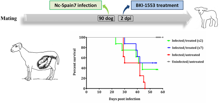
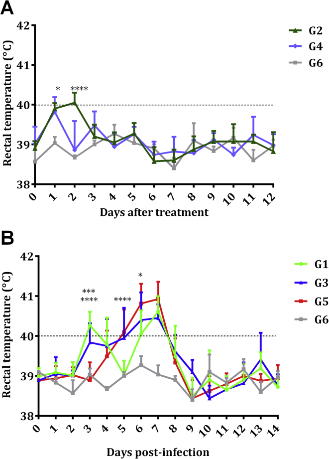
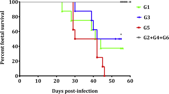
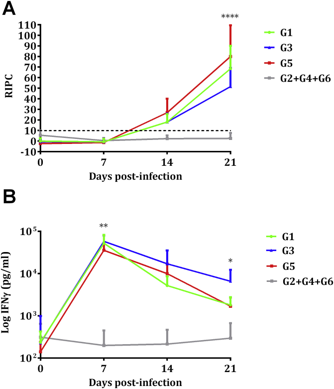
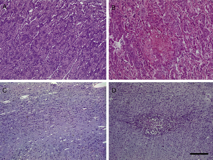
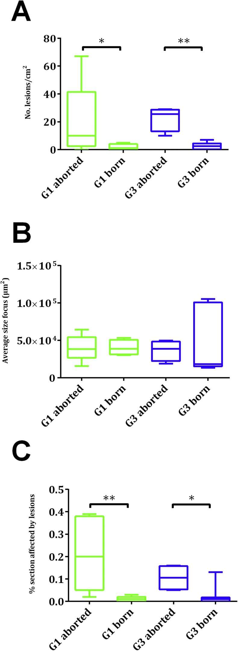
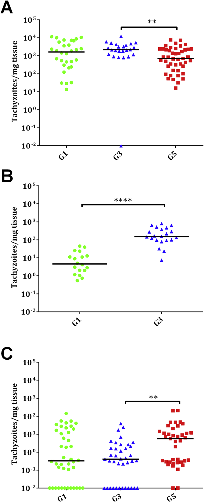
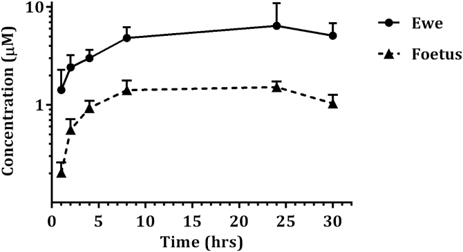
Neospora caninum is one of the main causes of abortion in cattle, and recent studies have highlighted its relevance as an abortifacient in small ruminants. Vaccines or drugs for the control of neosporosis are lacking. Bumped kinase inhibitors (BKIs), which are ATP-competitive inhibitors of calcium dependent protein kinase 1 (CDPK1), were shown to be highly efficacious against several apicomplexan parasites in vitro and in laboratory animal models. We here present the pharmacokinetics, safety and efficacy of BKI-1553 in pregnant ewes and foetuses using a pregnant sheep model of N. caninum infection. BKI-1553 showed exposure in pregnant ewes with trough concentrations of approximately 4 μM, and of 1 μM in foetuses. Subcutaneous BKI-1553 administration increased rectal temperatures shortly after treatment, and resulted in dermal nodules triggering a slight monocytosis after repeated doses at short intervals. BKI-1553 treatment decreased fever in infected pregnant ewes already after two applications, resulted in a 37-50% reduction in foetal mortality, and modulated immune responses; IFNγ levels were increased early after infection and IgG levels were reduced subsequently. N. caninum was abundantly found in placental tissues; however, parasite detection in foetal brain tissue decreased from 94% in the infected/untreated group to 69-71% in the treated groups. In summary, BKI-1553 confers partial protection against abortion in a ruminant experimental model of N. caninum infection during pregnancy. In addition, reduced parasite detection, parasite load and lesions in foetal brains were observed.

Keywords: BKI-1553; Neospora caninum; Pregnancy; Protein kinase inhibitor; Sheep; Treatment.


Figures

Image 1
Graphical abstract
Fig. 1
Fig. 1
Rectal temperatures of uninfected groups G2, G4 and G6 (A) and infected groups G1, G3 and G5 and the uninfected/untreated group G6 (B). Each point represents the mean + S.D. for each group. Rectal temperatures represented in the figure were analysed using Two-way ANOVA of repeated measures. For significant differences, (*) indicates P < 0.05, (***) indicates P < 0.001 and (****) indicates P < 0.0001.
Fig. 2
Fig. 2
Kaplan–Meier survival curves for foetuses in the infected groups G1 and G3 treated with BKI-1553, infected group G5, and the non-infected groups. Each point represents the percentage of surviving animals at that day, and downward steps correspond with observed deaths. Foetal survival curves were compared by the Log-rank (Mantel-Cox) test. For significant differences between foetal survival curves of infected groups, (*) indicates P < 0.05.
Fig. 3
Fig. 3
IgG response in sera (A) and IFNγ in supernatants of peripheral blood cell cultures (B). Values from infected (G1, G3 and G5) and uninfected (G2+G4+G6) pregnant ewes are represented. Each point represents the mean + S.D. at the different sampling times for each group. Data beyond day 21 pi are not shown, since several animals did not maintain pregnancy and were therefore sacrificed. Sera levels of total IgG antibodies against N. caninum are expressed as a relative index percent (RIPC), according to the formula: RIPC = (OD405 sample – OD405 negative control)/(OD405 positive control – OD405 negative control) × 100. Concentrations of IFNγ are expressed in pg/mL. Humoral and cellular immune responses represented in the figure were analysed using two-way ANOVA of repeated measures. For significant differences between infected groups, (*) indicates P < 0.05, (**) indicates P < 0.01 and (****) indicates P < 0.0001.
Fig. 4
Fig. 4
Hematoxylin and eosin staining. A) Interdigitate area of the placentome of an uninfected sheep with no evident lesion. B) Foci of necrosis and scant inflammatory infiltration at the interdigitated area of the placentome of an infected sheep. C) Foetal brain from an uninfected sheep with no evident lesion. D) Glial foci with central necrosis at the foetal brain from an infected sheep. Bar: 200 μm.
Fig. 5
Fig. 5
Box-plots showing number of lesions (A), average size of focus (B) and lesion rates (C) in foetal brains from G1 and G3. Graphs represent the median percentage, the lower and upper quartiles (boxes) and minimum and maximum values (whiskers). Histological measurements of lesions were analysed using the non-parametric Kruskal–Wallis test followed by Dunn's test for comparisons between groups, as well as the Mann–Whitney test for pairwise comparisons. For significant differences between infected and treated groups, (*) indicates P < 0.05 and (**) indicates P < 0.01.
Fig. 6
Fig. 6
Dot-plot graphs of N. caninum burdens in placentomes (A), cotyledons (B) and foetal brain (C) from G1, G3 and G5. Each dot represents individual values of parasite burden (number of parasites per mg of host tissue), and medians are represented as horizontal lines. Considering that the N. caninum detection limit by real-time PCR is 0.1 parasites, negative samples (0 parasites) were represented on the log scale as <0.1 (i.e., 10−2). Parasite burdens were analysed using the non-parametric Kruskal–Wallis test followed by Dunn's test for comparisons between groups, as well as the Mann–Whitney test for pairwise comparisons. For significant differences between infected groups in each tissue, (**) indicates P < 0.01 and (****) indicates P < 0.0001.
Fig. 7
Fig. 7
Plasma concentrations of BKI-1553 in pregnant ewes and foetuses after single 10 mg/kg subcutaneous dose. Each point represents the mean + S.D. at the different sampling times for pregnant ewes and foetuses.

Similar articles

Cited by

References

    1. Almería S., Serrano-Pérez B., Darwich L., Domingo M., Mur-Novales R., Regidor-Cerrillo J., Cabezón O., Pérez-Maillo M., López-Helguera I., Fernández-Aguilar X. Foetal death in naive heifers inoculated with Neospora caninum isolate Nc-Spain7 at 110 days of pregnancy. Exp. Parasitol. 2016;168:62–69. - PubMed
    1. Álvarez-García G., Collantes-Fernández E., Costas E., Rebordosa X., Ortega-Mora L.M. Influence of age and purpose for testing on the cut-off selection of serological methods in bovine neosporosis. Vet. Res. 2003;34:341–352. - PubMed
    1. Arranz-Solís D., Benavides J., Regidor-Cerrillo J., Horcajo P., Castaño P., del Carmen Ferreras M., Jiménez-Pelayo L., Collantes-Fernández E., Ferre I., Hemphill A., Pérez V., Ortega-Mora L.M. Systemic and local immune responses in sheep after Neospora caninum experimental infection at early, mid and late gestation. Vet. Res. 2016;47:1–13. - PMC - PubMed
    1. Arranz-Solís D., Benavides J., Regidor-Cerrillo J., Fuertes M., Ferre I., Ferreras Mdel C., Collantes-Fernandez E., Hemphill A., Perez V., Ortega-Mora L.M. Influence of the gestational stage on the clinical course, lesional development and parasite distribution in experimental ovine neosporosis. Vet. Res. 2015;46:19. - PMC - PubMed
    1. Buxton D., Blewett D., Trees A., McColgan C., Finlayson J. Further studies in the use of monensin in the control of experimental ovine toxoplasmosis. J. Comp. Pathol. 1988;98:225–236. - PubMed

Publication types

MeSH terms