

A rare case of hydrometrocolpos from persistent urogenital sinus in patient affected by adrenogenital syndrome
- PMID: 29502244
- PMCID: PMC6113191
- DOI: 10.1007/s40477-018-0290-9
A rare case of hydrometrocolpos from persistent urogenital sinus in patient affected by adrenogenital syndrome
Abstract
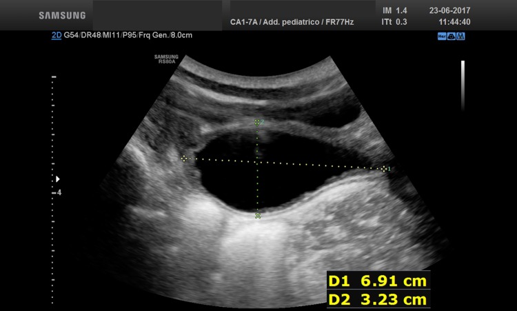
Persistent urogenital sinus (PUGS) is a congenital pathological condition characterized by an abnormal communication between the urethra and vagina, which has an estimated incidence of 0.6/10,000 female births. It could be the only known malformation or part of a syndrome. PUGS is commonly shown by a pelvic mass, related to a distended bladder, hydrometrocolpos which is due to an obstruction leading to the dilation of the vagina and uterus (i.e., imperforate hymen, transverse vaginal septum or atresia, and PUGS) or both. We present a case of female patient with classical congenital adrenal hyperplasia, diagnosed on the 7th day of life, with ambiguous genitalia, untreated surgically only with hormone therapy by parental decision. The patient, at the age of 5 years and 5 months, came to our observation for a pelvic ultrasound, which revealed retrovesical neoformation with anechoic content and regular walls. We performed the ultrasound examination that showed the dilation of the cervix and the vaginal canal with anechoic finely corpuscolated content in the declining portion, compatible with hydrometrocolpos from probable persistence of the urogenital sinus. The voiding cystourethrography (VCUG) confirmed the ultrasound diagnosis, with evidence of urogenital sinus. In conclusion, ultrasound is the first diagnostic tool, but need to be completed by other technical procedures, which VCUG or magnetic resonance imaging to observe the site of fusion of the urinary and genital tract.
La persistenza del seno urogenitale (PUGS) è una rara condizione patologica congenita, caratterizzata da un’anomala comunicazione tra uretra e vagina, la cui incidenza è di 0.6/10.000 neonate. Clinicamente, può rappresentare l’unica malformazione rilevabile o far parte di un quadro sindromico. Si presenta comunemente come una massa pelvica, associata a distensione vescicale e/o idrometrocolpo, ossia dilatazione della vagina e dell’utero causata dall’accumulo di secrezioni cervicali conseguente ad un’ostruzione (imene imperforato, setto vaginale trasverso, atresia vaginale e PGUS). Viene presentato il caso di una paziente di sesso femminile, affetta da Sindrome Adreno-Genitale (SAG) forma classica, diagnosticata al 7° giorno di vita, con genitali ambigui (ipertrofia clitoridea, grandi labbra scrotalizzate) non trattati chirurgicamente per decisione parentale, in terapia ormonale sostitutiva. La paziente, all’età di 5 anni e 5 mesi, giunge alla nostra osservazione per una richiesta di ecografia della regione pelvica richiesta, come consulenza, per una neoformazione retrovescicale a contenuto anecogeno e pareti regolari. L’esame ecografico ha mostrato una dilatazione del collo e del canale vaginale a contenuto anecogeno finemente corpuscolato nella porzione declive, compatibile con idrometrocolpo da verosimile persistenza del seno urogenitale. La cistouretrografia ha confermato il reperto ecografico, con evidenza del seno uro-genitale. In conclusione, l’ecografia è la metodica di prima istanza utilizzata, tuttavia necessita di ulteriori metodiche, quali la Cistouretrografia e/o la Risonanza Magnetica per rilevare il punto di comunicazione tra il tratto urinario ed il tratto genitale.
Keywords: Hydrometrocolpos; Magnetic resonance imaging (MRI); Persistent urogenital sinus (PUGS); Ultrasound (US); Voiding cystourethrography (VCUG).
Conflict of interest statement
Conflict of interest
The authors declare that they have no conflict of interest.
Ethical approval
All procedures performed in studies involving human participants were in accordance with the ethical standards of the institutional and/or national research committee and with the 1964 Helsinki declaration and its later amendments or comparable ethical standards.
Human and animal rights
This article does not contain any studies with animals performed by any of the authors.
Informed consent
Informed consent was obtained from all individual participants included in the study.
Figures


Similar articles
-
Hydrometrocolpos: a Contemporary Review of the Last 5 Years.Curr Urol Rep. 2023 Dec;24(12):601-610. doi: 10.1007/s11934-023-01191-4. Epub 2023 Dec 1. Curr Urol Rep. 2023. PMID: 38038828 Review.
-
Antenatal MR diagnosis of urinary hydrometrocolpos due to urogenital sinus.Pediatr Radiol. 2006 Oct;36(10):1086-9. doi: 10.1007/s00247-006-0249-4. Epub 2006 Jun 30. Pediatr Radiol. 2006. PMID: 16810498
-
Persistent Urogenital Sinus: Diagnostic Imaging for Clinical Management. What Does the Radiologist Need to Know?Am J Perinatol. 2016 Apr;33(5):425-32. doi: 10.1055/s-0035-1565996. Epub 2015 Oct 21. Am J Perinatol. 2016. PMID: 26489064 Review.
-
Fetal hydrometrocolpos, uterus didelphys with low vaginal and anal atresia: difficulties in differentiation from a complex cloacal malformation: a case report.Genet Couns. 2012;23(4):513-7. Genet Couns. 2012. PMID: 23431753
-
Fetal urogenital sinus with consecutive hydrometrocolpos because of labial fusion: prenatal diagnostic difficulties and postpartal therapeutic management.Fetal Diagn Ther. 2008;23(4):287-92. doi: 10.1159/000123615. Epub 2008 Apr 14. Fetal Diagn Ther. 2008. PMID: 18417994
Cited by
-
Hydrometrocolpos: a Contemporary Review of the Last 5 Years.Curr Urol Rep. 2023 Dec;24(12):601-610. doi: 10.1007/s11934-023-01191-4. Epub 2023 Dec 1. Curr Urol Rep. 2023. PMID: 38038828 Review.
-
Acute epiploic appendagitis: ultrasound and computed tomography findings of a rare case of acute abdominal pain and the role of other imaging techniques.Pol J Radiol. 2020 Apr 6;85:e178-e182. doi: 10.5114/pjr.2020.94335. eCollection 2020. Pol J Radiol. 2020. PMID: 32419882 Free PMC article.
References
-
- Valentini AL, Giuliani M, Gui B, Laino M, Zecchi V, Rodolfino E, Ninivaggi V, Manzoni C, Bonomo L. Persistent urogenital sinus: diagnostic imaging for clinical management. What does the radiologist need to know? Am J Perinatol. 2016;33:425–432. - PubMed
Publication types
MeSH terms
Supplementary concepts
LinkOut - more resources
Full Text Sources
Other Literature Sources
Medical
