. 2018 Mar 13;10(3):751-765.
doi: 10.1016/j.stemcr.2018.01.041. Epub 2018 Mar 1.

Renal Subcapsular Transplantation of PSC-Derived Kidney Organoids Induces Neo-vasculogenesis and Significant Glomerular and Tubular Maturation In Vivo

Affiliations

Renal Subcapsular Transplantation of PSC-Derived Kidney Organoids Induces Neo-vasculogenesis and Significant Glomerular and Tubular Maturation In Vivo

Cathelijne W van den Berg et al. Stem Cell Reports. .

Abstract

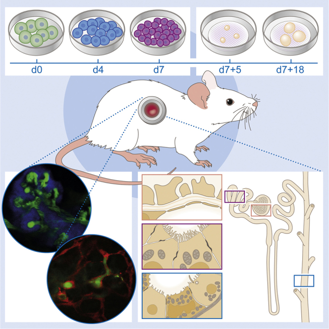
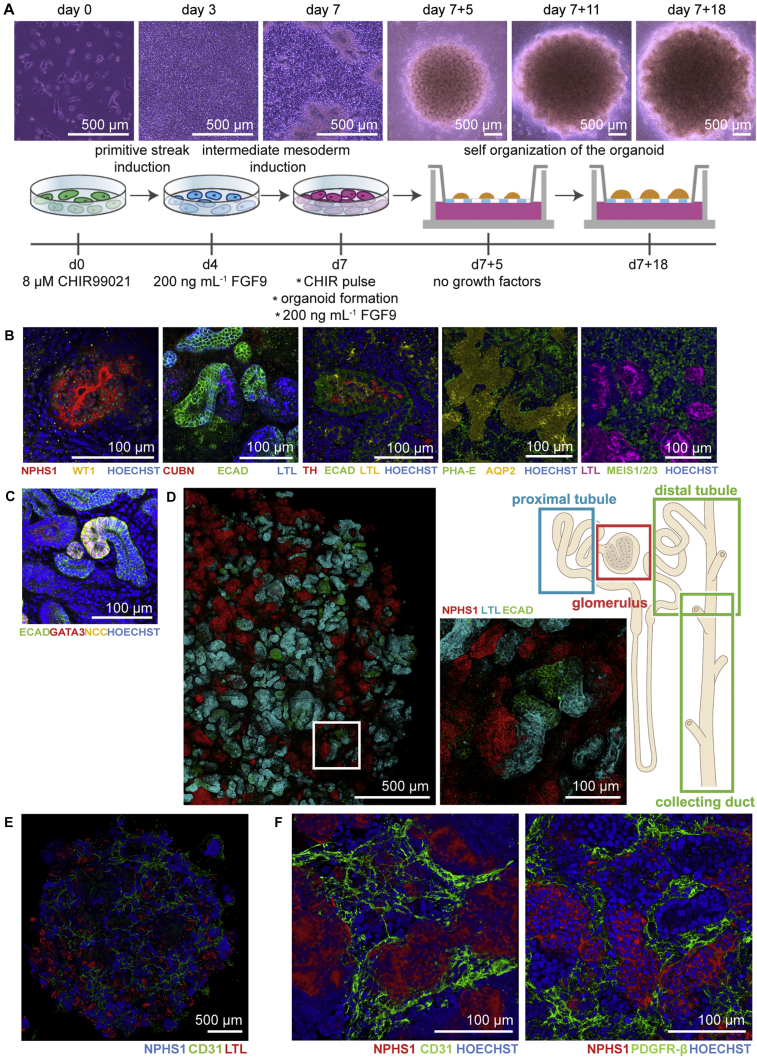
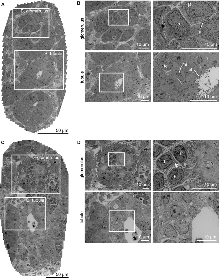
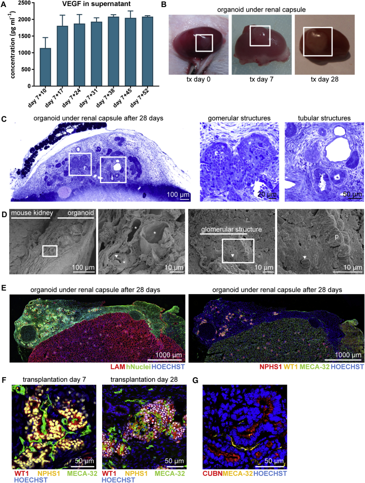
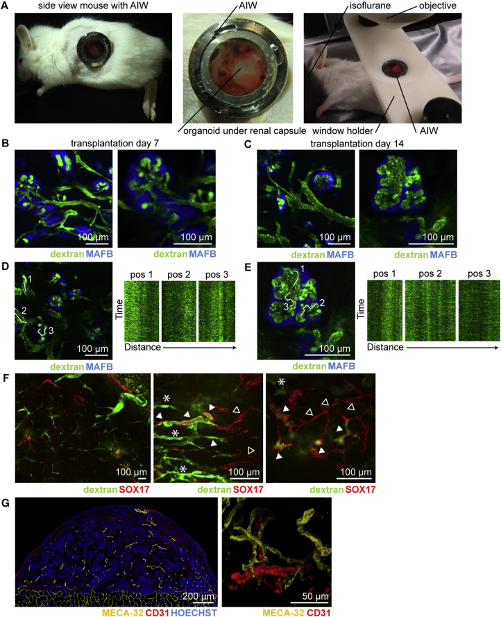
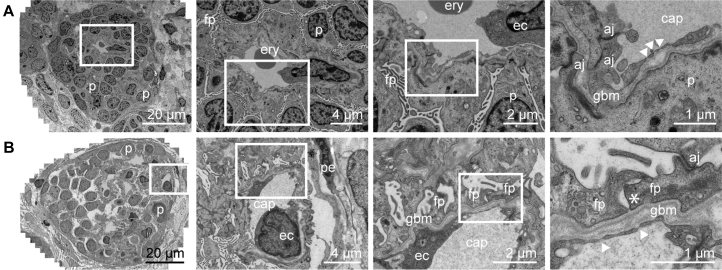
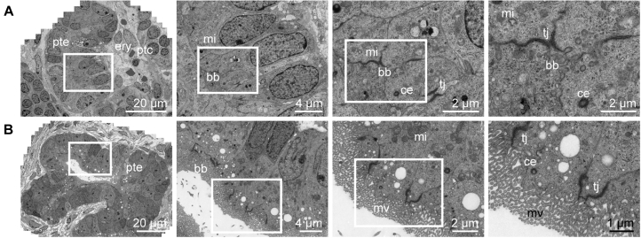
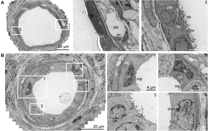
Human pluripotent stem cell (hPSC)-derived kidney organoids may facilitate disease modeling and the generation of tissue for renal replacement. Long-term application, however, will require transferability between hPSC lines and significant improvements in organ maturation. A key question is whether time or a patent vasculature is required for ongoing morphogenesis. Here, we show that hPSC-derived kidney organoids, derived in fully defined medium conditions and in the absence of any exogenous vascular endothelial growth factor, develop host-derived vascularization. In vivo imaging of organoids under the kidney capsule confirms functional glomerular perfusion as well as connection to pre-existing vascular networks in the organoids. Wide-field electron microscopy demonstrates that transplantation results in formation of a glomerular basement membrane, fenestrated endothelial cells, and podocyte foot processes. Furthermore, compared with non-transplanted organoids, polarization and segmental specialization of tubular epithelium are observed. These data demonstrate that functional vascularization is required for progressive morphogenesis of human kidney organoids.

Keywords: directed differentiation; human pluripotent stem cells; intravital microscopy; kidney organoids; maturation; transplantation; vascularization.


Figures

None
Graphical abstract
Figure 1
Figure 1
Kidney Organoids Derived from Induced Pluripotent Stem Cells Display Structures Characteristic of a Nephron on Day 7 + 18 of Differentiation (A) Bright-field images and schematic of the experimental time line for the generation of kidney organoids. (B) Immunofluorescence analysis of the different segments of the nephron: podocytes (WT1+, NPHS1+), proximal tubule (CUBN+, PHA-E+, LTL+), Lis of Henle (Tamm Horsfall+), distal tubule (ECAD+), collecting duct (AQP-2+), and stromal cells (MEIS1/2/3+) visualized as 3D structures. (C) Presence of the sodium chloride (NCC) symporter in the kidney organoid. (D) Tile scan of an immunofluorescent organoid demonstrating anatomically correct interconnected segments of the nephron: glomerulus, proximal tubule, distal tubule, and collecting duct. Close-up view of the boxed area displays the structures in detail. (E) Endothelial cells (CD31+) are mainly localized around glomerular structures (NPHS1+) and not around tubular structures (LTL+). (F) The CD31+ cells do not invade glomerular structures (NPHS1+) and pericytes (PDGFR-β+) surround glomerular structures.
Figure 2
Figure 2
Ultrastructural Evaluation Shows Lack of Advanced Maturation of Kidney Organoids In Vitro over Time at Day 7 + 18 and Day 7 + 53 (A) Low-magnification transmission electron microscopy (TEM) tile scan of kidney organoid cultured for 7 + 18 days on the air-liquid interface displaying glomerular and tubular structures. (B) High-magnification TEM images of boxed areas in (A) demonstrate characteristic structures, such as podocytes with primitive foot processes in glomerulus (top) and brush border with microvilli in the open lumen of a tubular structure (bottom). (C) Low-magnification TEM image of a kidney organoid cultured for prolonged time (7 + 53 days) on the air-liquid interface. (D) Detailed TEM images of boxed areas in (C) from glomerular structure (top) and tubular structure (bottom) demonstrate features of maturation, such as foot processes, formation of glomerular basement membrane, and microvilli, respectively. However, the structures are more disorganized. p, podocyte; fp, foot processes; te, tubular epithelium; mi, mitochondria; bb, brush border.
Figure 3
Figure 3
Kidney Organoids Become Vascularized upon Transplantation for 7 and 28 Days (A) Concentration of VEGF (pg mL−1) determined by Luminex assay in the supernatant of three cultured organoids measured weekly from day 7 + 10 until day 7 + 52. Data are represented as means ± SEM. (B) Transplanted human kidney organoid under renal capsule of mice on the day of transplantation (tx), after 7 and 28 days showing growth upon vascularization. (C) Toluidine blue staining of organoid under the renal capsule after 28 days of transplantation. Boxed areas highlight glomerular and tubular structures displayed on the right. (D) Scanning electron microscopy images suggests blood vessels in the kidney organoid after transplantation and inside a glomerular structure. Close-up views of boxed areas are displayed. (E) Immunofluorescent overview of human nuclei and LAMININ (LAM) in the organoid under the renal capsule of a mouse kidney (left) and integration of mouse endothelial cells (MECA-32+) in the organoid and glomerular structures (right). (F) Mouse endothelial cells (MECA-32+) were observed in association with glomerular structures (NPHS1+, WT1+) in the human kidney organoid after 7 and 28 days of transplantation. (G) Peritubular vascularization observed as MECA-32+ endothelial cells aligning tubular (CUBN+) structures. p, podocyte; arrowhead, erythrocyte; asterisk, blood vessels.
Figure 4
Figure 4
In Vivo Imaging of Vascularized Kidney Organoids Shows Glomerular Vascularization and Chimeric Organoid Circulation after 7 and 14 Days of Transplantation (A) Mouse with the abdominal imaging window (AIW) on the left kidney and a close-up view of a kidney window after 14 days of implantation. Right picture shows the mouse with the AIW installed for microscope analysis. (B and C) In vivo imaging of transplanted organoids derived from hiPSC-MAFB-BFP after 7 days (B) and 14 days (C) of transplantation. Circulating plasma was visualized with 2,000 kDa FITC-labelled dextran and flow was detected in BFP+ glomerular structures. (D and E) Kymographs from marked positions demonstrating the dynamic blood flow through the capillary in the organoid (D) and inside the glomerular structure (E) after 14 days of transplantation. (F) Low and high magnification of hESC-SOX17-mCherry derived kidney organoids after transplantation for 14 days revealing mCherry+ endothelial cells perfused with FITC-labelled dextran (arrowhead) and host-derived vasculature (asterisk). Note that not all mCherry+ endothelial cells were perfused (open arrowhead). (G) Immunofluorescence of human CD31+ and mouse MECA-32+ in the transplanted kidney organoid.
Figure 5
Figure 5
Wide-Field Ultrastructural Evaluation of Transplanted Organoids Shows Evidence for Glomerular Vascularization and Maturation Transmission electron micrographs of vascularized glomerular structures after 7 days (A) and 28 days (B) of transplantation. Glomeruli become organized displaying features of maturation such as foot processes, formation of glomerular basement membrane and progression toward the formation of a slit diaphragm. Close-up views of the boxed areas are displayed. p, podocyte; fp, foot processes; ery, erythrocyte; ec, endothelial cell; aj, adherens junctions; gbm, glomerular basement membrane; arrowhead, endothelial fenestrae; cap, capillary; pe, parietal epithelium; asterisk, developing slit diaphragm.
Figure 6
Figure 6
Wide-Field Ultrastructural Evaluation of Transplanted Organoids Shows Advanced Tubular Epithelial Differentiation Transmission electron micrographs of developing tubular structure with peritubular capillaries after 7 days (A) and 28 days (B) of transplantation under the renal capsule. After transplantation, the tubular structures display a single layer of epithelial cells connected with tight junctions, have an open lumen and show tubular polarization, such as characteristic mitochondria, microvilli, and centrioles. Close-up views of the boxed areas are displayed. pte, proximal tubular epithelium; ptc, peritubular capillary; ery, erythrocyte; mi, mitochondria; bb, brush border; tj, tight junction; ce, centriole; mv, microvilli.
Figure 7
Figure 7
Wide-Field Ultrastructural Evaluation of Transplanted Organoids Demonstrates Peritubular Vascularization and Intra-tubular Specification (A) Transmission electron microscopy stitch showing tubular structures after 28 days of transplantation displaying an open lumen with a single layer of epithelial cells. The tubule has a brush border, tight junctions, and a peritubular capillary. (B) Transmission electron micrographs showing tubular dilation with micro projections that suggest the presence of intercalated cells existing in the collecting duct-type structure. Boxed areas correspond with numbered close-up views. te, tubular epithelium, ptc, peritubular capillary; bb, brush border; ec, endothelial cell; tj, tight junction; ic, intercalated cell; ci, cilium; ce, centriole; mv, microvilli; mi, mitochondria; mp, micro projections.

References

    1. Barri Y.M., Munshi N.C., Sukumalchantra S., Abulezz S.R., Bonsib S.M., Wallach J., Walker P.D. Podocyte injury associated glomerulopathies induced by pamidronate. Kidney Int. 2004;65:634–641. - PubMed
    1. Briggs J.A., Sun J., Shepherd J., Ovchinnikov D.A., Chung T.L., Nayler S.P., Kao L.P., Morrow C.A., Thakar N.Y., Soo S.Y. Integration-free induced pluripotent stem cells model genetic and neural developmental features of down syndrome etiology. Stem Cells. 2013;31:467–478. - PubMed
    1. Chen G., Gulbranson D.R., Hou Z., Bolin J.M., Ruotti V., Probasco M.D., Smuga-Otto K., Howden S.E., Diol N.R., Propson N.E. Chemically defined conditions for human iPSC derivation and culture. Nat. Methods. 2011;8:424–429. - PMC - PubMed
    1. Costa M., Dottori M., Ng E., Hawes S.M., Sourris K., Jamshidi P., Pera M.F., Elefanty A.G., Stanley E.G. The hESC line Envy expresses high levels of GFP in all differentiated progeny. Nat. Methods. 2005;2:259–260. - PubMed
    1. Costa M., Sourris K., Hatzistavrou T., Elefanty A.G., Stanley E.G. Expansion of human embryonic stem cells in vitro. Curr. Protoc. Stem Cell Biol. 2008;Chapter 1 Unit 1C.1.1–1C.1.7. - PubMed

Publication types