. 2018 Mar 13;10(3):739-750.
doi: 10.1016/j.stemcr.2018.01.040. Epub 2018 Mar 1.

Functional Beta Cell Mass from Device-Encapsulated hESC-Derived Pancreatic Endoderm Achieving Metabolic Control

Affiliations

Functional Beta Cell Mass from Device-Encapsulated hESC-Derived Pancreatic Endoderm Achieving Metabolic Control

Thomas Robert et al. Stem Cell Reports. .

Abstract

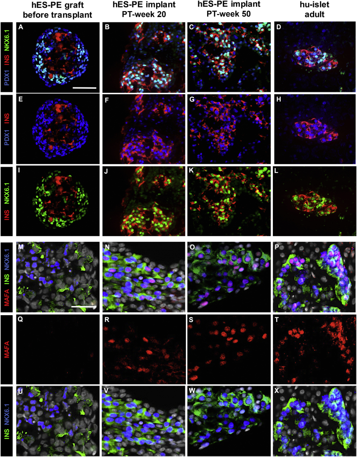
Human stem cells represent a potential source for implants that replace the depleted functional beta cell mass (FBM) in diabetes patients. Human embryonic stem cell-derived pancreatic endoderm (hES-PE) can generate implants with glucose-responsive beta cells capable of reducing hyperglycemia in mice. This study with device-encapsulated hES-PE (4 × 10⁶ cells/mouse) determines the biologic characteristics at which implants establish metabolic control during a 50-week follow-up. A metabolically adequate FBM was achieved by (1) formation of a sufficient beta cell number (>0.3 × 10⁶/mouse) at >50% endocrine purity and (2) their maturation to a functional state comparable with human pancreatic beta cells, as judged by their secretory responses during perifusion, their content in typical secretory vesicles, and their nuclear NKX6.1-PDX1-MAFA co-expression. Assessment of FBM in implants and its correlation with in vivo metabolic markers will guide clinical translation of stem cell-derived grafts in diabetes.

Keywords: diabetes; differentiation; encapsulation; functional beta cell mass; functional maturation; metabolic control; stem cell therapy; stem cell-derived pancreatic endoderm.


Figures

None
Graphical abstract
Figure 1
Figure 1
Development of FBM in Device-Encapsulated hES-PE Implants Followed over 50 Weeks (A) Plasma hu-C-peptide (15 min after intraperitoneal glucose load) and glucagon levels (basal, 2 hr fast) (means ± SD) in NSG-recipient mice (filled squares, n = 20) increased during the first 20 weeks as in NOD/SCID recipients (filled circles, n = 19), the strain also used in our previous study (Motté et al., 2014). NOD/SCID control mice (n = 9) are plotted as empty circles. Plasma hu-C-peptide became consistently detectable from PT week 10 onward, and increased in all animals to levels stabilizing between weeks 30 and 50. Plasma hu-C-peptide levels are also shown for NOD/SCID recipients of human pancreatic islet cells (106 beta cells/recipient) under the kidney capsule (triangles, dotted line); they were significantly higher than values in hES-PE recipients at PT weeks 5 and 10 (∗∗∗p < 0.0001 and p < 0.05 by one-way ANOVA with Tukey's post-hoc test, respectively), but became lower at later time points. Plasma glucagon in NSG recipients was higher than in controls (empty squares, n = 7) from PT weeks 7 to 32 (p < 0.05; ∗∗p < 0.01; ∗∗∗p < 0.001 by one-way ANOVA with Tukey's post-hoc test); after which the difference was no longer statistically significant. (B) At PT week 50, plasma hu-C-peptide levels correlated with the number of beta cells and the number of alpha cells in the retrieved implants (linear regression with 95% confidence interval of, respectively, rp = 0.9555; R2 = 0.9130; p = 0.0002, and rp = 0.9857; R2 = 0.9716; p < 0.0001).
Figure 2
Figure 2
Glucose Responsiveness of Insulin Release by hES-PE Implants Retrieved at PT Week 50 Data express means ± SD of insulin release by hES-PE implants retrieved at PT week 50 (black curve, n = 8) and by cultured human pancreatic islet cell isolates (gray curve, n = 7). Both preparations were perifused at varying glucose concentrations. The response to 20 mmol/L glucose was first measured in the absence and then in the presence of glucagon (10−8 mol/L). Insulin was measured in the perifusate fractions and expressed per 103 beta cells (top) or as percent of cellular insulin content (bottom).
Figure 3
Figure 3
Expression of Beta Cell-Specific Transcription Factors in Insulin-Positive Cells of hES-PE Implants (A–L) The hES-PE aggregates in the graft exhibited peripherally located INS-negative cells with nuclear co-expression of NKX6.1 and PDX1, whereas centrally located INS-positive cells were negative for these transcription factors (A, E, and I). At PT weeks 20 and 50 virtually all INS-positive cells presented nuclear positivity for PDX1 (B, C, F, and G) and NKX6.1 (B, C, J, and K), as in human pancreatic beta cells (D, H, and L). Scale bar, 50 μm. (M–X) Nuclear expression of MAFA was absent in hES-PE at the start (M, Q, and U), but clearly present at PT weeks 20 and 50 and restricted to insulin-positive cells. The percentage of MAFA + INS + cells increased from 25% ± 8% at PT week 20 (N, R, and V) to 53% ± 21% at PT week 50 (p < 0.01) (O, S, and W), approaching that in the adult human pancreas (P, T, and X) (71% ± 18%). Scale bar, 25 μm.
Figure 4
Figure 4
Metabolic Effects in Subgroup of Recipients with hu-C-Peptide Levels >6 ng/mL from PT Weeks 20 to 50 Basal glycemia and plasma mouse C-peptide levels decreased under levels measured at PT week 0 (°°p < 0.01; °°°p < 0.001) and in controls (empty squares, n = 4–7, p < 0.05; ∗∗p < 0.01; ∗∗∗p < 0.001). Both parameters were also lower following intraperitoneal glucose load (statistical difference with PT week 5: °p < 0.05; °°°p < 0.001, and with controls: ∗∗∗p < 0.001). Values are expressed as means ± SD.

References

    1. Agulnick A.D., Ambruzs D.M., Moorman M.A., Bhoumik A., Cesario R.M., Payne J.K., Kelly J.R., Haakmeester C., Srijemac R., Wilson A.Z. Insulin-producing endocrine cells differentiated in vitro from human embryonic stem cells function in macroencapsulation devices in vivo. Stem Cells Transl. Med. 2015;4:1214–1222. - PMC - PubMed
    1. Bouwens L., Wang R.N., De Blay E., Pipeleers D.G., Kloppel G. Cytokeratins as markers of ductal cell differentiation and islet neogenesis in the neonatal rat pancreas. Diabetes. 1994;43:1279–1283. - PubMed
    1. Bruin J.E., Rezania A., Xu J., Narayan K., Fox J.K., O'Neil J.J., Kieffer T.J. Maturation and function of human embryonic stem cell-derived pancreatic progenitors in macroencapsulation devices following transplant into mice. Diabetologia. 2013;56:1987–1998. - PubMed
    1. D'Amour K.A., Agulnick A.D., Eliazer S., Kelly O.G., Kroon E., Baetge E.E. Efficient differentiation of human embryonic stem cells to definitive endoderm. Nat. Biotechnol. 2005;23:1534–1541. - PubMed
    1. D'Amour K.A., Bang A.G., Eliazer S., Kelly O.G., Agulnick A.D., Smart N.G., Moorman M.A., Kroon E., Carpenter M.K., Baetge E.E. Production of pancreatic hormone-expressing endocrine cells from human embryonic stem cells. Nat. Biotechnol. 2006;24:1392–1401. - PubMed

Publication types