. 2018 Apr 4;26(4):1040-1055.
doi: 10.1016/j.ymthe.2018.01.011. Epub 2018 Jan 31.

Local Delivery of miR-21 Stabilizes Fibrous Caps in Vulnerable Atherosclerotic Lesions

Affiliations

Local Delivery of miR-21 Stabilizes Fibrous Caps in Vulnerable Atherosclerotic Lesions

Hong Jin et al. Mol Ther. .

Abstract

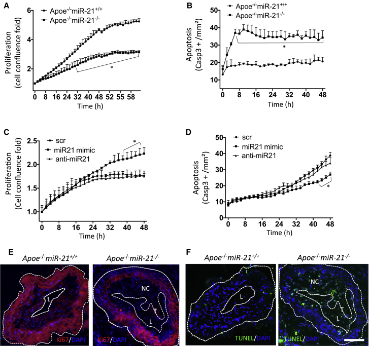
miRNAs are potential regulators of carotid artery stenosis and concordant vulnerable atherosclerotic plaques. Hence, we analyzed miRNA expression in laser captured micro-dissected fibrous caps of either ruptured or stable plaques (n = 10 each), discovering that miR-21 was significantly downregulated in unstable lesions. To functionally evaluate miR-21 in plaque vulnerability, miR-21 and miR-21/apolipoprotein-E double-deficient mice (Apoe-/-miR-21-/-) were assessed. miR-21-/- mice lacked sufficient smooth muscle cell proliferation in response to carotid ligation injury. When exposing Apoe-/-miR-21-/- mice to an inducible plaque rupture model, they presented with more atherothrombotic events (93%) compared with miR-21+/+Apoe-/- mice (57%). We discovered that smooth muscle cell fate in experimentally induced advanced lesions is steered via a REST-miR-21-REST feedback signaling pathway. Furthermore, Apoe-/-miR-21-/- mice presented with more pronounced atherosclerotic lesions, greater foam cell formation, and substantially higher levels of arterial macrophage infiltration. Local delivery of a miR-21 mimic using ultrasound-targeted microbubbles into carotid plaques rescued the vulnerable plaque rupture phenotype. In the present study, we identify miR-21 as a key modulator of pathologic processes in advanced atherosclerosis. Targeted, lesion site-specific overexpression of miR-21 can stabilize vulnerable plaques.

Keywords: atherosclerosis; microRNA; molecular medicine.


Figures

None
Graphical abstract
Figure 1
Figure 1
Decreased miR-21 Expression Is Associated with Atherosclerotic Fibrous Cap Instability and Rupture (A) Staining for laser captured fibrous caps of ten ruptured versus ten stable human carotid atherosclerotic plaques. FC, fibrous cap; L, lumen; NC, necrotic core; T, thrombus. (B) qRT-PCR analysis shows significant miR-21 downregulation in ruptured versus stable plaques (mean + SEM). Groups were compared using Student’s t test. (C) In situ hybridization (ISH) of human carotid arteries indicate that miR-21 is expressed in the intima-media, as well as fibrous cap structures of stable plaques, whereas it appears reduced in ruptured lesions. *p < 0.05 versus stable. Bar, 100 μm.
Figure 2
Figure 2
miR-21 Deficiency Suppresses Neointima Formation and Fibrous Cap Stability in Mouse Models of Advanced Atherosclerosis (A) Histologic images stained with H&E and smooth muscle cell α-actin (SMA) of the right common carotid artery (300 μm from the site of complete ligation). (B) Descriptive cartoon of the mouse carotid plaque rupture model. Male Apoe-deficient mice, 12 weeks of age, receive an incomplete carotid artery ligation directly below the bifurcation. Within 4 weeks, a plaque forms proximal to the ligation. Placement of a cast with a conically shaped internal lumen results in plaque rupture in about 50%–60% of Apoe−/− mice. (C) ISH of right carotid arteries from Apoe-deficient mice shows significantly decreased miR-21 expression in ruptured versus stable plaques. (D) Histologic images stained with H&E, α-SMA, and cross-linked fibrin of stable and ruptured carotid plaques in Apoe−/−miR-21+/+ and Apoe−/−miR-21−/− mice. FC, fibrous cap; L, lumen; NC, necrotic core; T, thrombus. (E) Apoe−/−miR-21−/− mice (n = 15) display significantly higher plaque rupture rates compared with Apoe−/−miR-21+/+ mice (n = 14) (p = 0.02). Data were analyzed using chi-square or Fisher’s exact test. *p < 0.05 versus Apoe−/−miR-21+/+. Bar, 100 μm.
Figure 3
Figure 3
miR-21 Deficiency Limits SMC Proliferation While Enhancing Apoptosis (A) Substantially suppressed proliferation of cultured primary aortic SMCs from Apoe−/−miR-21−/− mice compared with cells from Apoe−/−miR-21+/+ littermate controls (n = 3 per group). Proliferation is indicated as percentage of confluence and plotted as fold increase (percentage confluence at specific time divided by percentage confluence at time point 0). (B) Enhanced apoptosis of primary aortic SMCs from Apoe−/−miR-21−/− mice. (C) miR-21 mimic treatment stimulates human carotid SMC proliferation significantly. (D) miR-21 mimic significantly inhibits HCtASMC apoptosis. (E) Ki-67 immunofluorescence staining confirms decreased proliferation in the carotid plaque of Apoe−/−miR-21−/− mice. L, lumen; NC, necrotic core. (F) Increased SMC apoptosis assessed in the carotid plaque of Apoe−/−miR-21−/− mice by using the TUNEL assay. *p < 0.05 versus Apoe−/−miR-21+/+ or SCR. Mean ± SEM. Bar, 100 μm. For (A)–(D), data were obtained from two experiments run in duplicates. For (E) and (F), data were obtained from two experiments run in triplicate. Data presented in (A)–(D) were analyzed using two-way repeated-measures ANOVA.
Figure 4
Figure 4
REST-miR-21 Feedback Loop Regulates SMC Fate (A) Mouse transcriptome array (MTA) profiling data shows significant upregulation of REST in advanced carotid artery plaques compared with uninjured control arteries; FDR p value is expressed as binary logarithm. (B) REST mRNA expression is significantly upregulated in advanced lesion material compared with control carotid artery. (C) Increased REST protein levels in ruptured carotid plaques of male Apoe-deficient mice assessed with REST IHC staining. L, lumen; NC, necrotic core. (D) REST IHC staining indicates higher REST levels in the carotid artery and plaques of Apoe−/−miR-21−/− mice. (E) REST overexpression with plasmid transfection suppresses proliferation in cultured HCtASMCs, while stimulating their apoptosis. REST knockdown using siRNA increases cell proliferation significantly. (F) Overexpression of REST leads to miR-21 reduction, while REST inhibition increases miR-21 levels in cultured human carotid artery SMCs. (G) miR-21 modulation using mimics and anti-miRs in cultured human carotid artery SMCs reversely regulates REST mRNA expression (n = 3 per group; the transfection experiments were repeated twice for E–G). (H) Predicted human-miR-21-5p binding site in the REST 3′UTR. (I) Luciferase reporter assay indicates REST as a direct target of mi-21-5p upon miR-21 mimic stimulation. (J) TNF-α increases REST expression levels in human carotid artery SMCs compared with control. Treatment with oxLDL has no significant effect. (K) REST mRNA expression significantly increased in ruptured versus stable atherosclerotic plaques from human patients with carotid artery disease. (L) Immunohistochemical-based staining for REST indicates elevated levels in ruptured versus stable human carotid artery plaques. *p < 0.05 versus NC or SCR or stable. Mean ± SEM. Bar, 100 μm. Data in (E) were analyzed using two-way repeated-measures ANOVA; all other data were compared using Student’s t test.
Figure 5
Figure 5
miR-21 Targets PTEN/AKT/ERK Signaling in SMCs (A) Human-miR-21-5p non-canonical binding site in the PTEN 3′UTR. (B–D) Significant PTEN mRNA (B) and protein reduction (C and D) after miR-21 mimic versus scramble treatment in human carotid artery SMCs. (E) Western immunoblotting confirms significantly increased levels of PTEN and decreased phosphorylated AKT and ERK1/2 in aortic tissue specimen of Apoe−/−miR-21−/− mice compared with those from their Apoe−/−miR-21+/+ littermate controls (n = 3 per group). (F) Apoe−/−miR-21−/− mice display higher PTEN expression in fibrous caps of advanced atherosclerotic plaques and carotid arteries upon PTEN immunofluorescent staining. FC, fibrous cap; L, lumen; T, thrombus. *p < 0.05 versus SCR. Mean ± SEM. Bar, 100 μm. Groups were compared using Student’s t test.
Figure 6
Figure 6
miR-21 Deficiency Activates NF-kB Nuclear Translocation and Enhances OxLDL Uptake in Mouse Peritoneal Macrophages (A and B) Atherosclerotic lesions were assessed by staining with Sudan IV (A) and expressed as percentage surface area for the entire aorta as well as individual segments (B). Data are given as mean ± SEM (n = 5; *p = 0.03 versus the Apoe−/−miR-21+/+ mice). (C) Substantially larger atherosclerotic lesions could be detected in aortic roots of Apoe−/−miR-21−/− mice assessed with oil red O staining. (D) Increased macrophage deposition and activity (Mac-2-positive cells) in carotid artery atherosclerotic plaques of Apoe−/−miR-21−/− mice. (E) Greatly enhanced FITC-conjugated oxLDL (green) uptake of peritoneal macrophages (Alexa Fluor 647 labeled red) of Apoe−/−miR-21−/− mice (n = 7), compared with Apoe−/−miR-21+/+-derived macrophages (n = 8), which could be effectively blocked via pretreatment with a NF-kB translocation inhibitor (indicated with arrows; Apoe−/−miR-21−/−, n = 5; Apoe−/−miR-21+/+, n = 3) being assessed kinetically with live-cell imaging (F). (G) Fluorescent staining with anti NF-kB p65, to determine localization (nuclear) in macrophages with or without oxLDL stimulation. (H) Quantification indicates increased levels of NF-kB nuclear translocation of Apoe−/−miR-21−/− mice macrophages before oxLDL stimulation, which becomes further enhanced upon stimulation with oxLDL (n = 4). (I) Supernatant of cultured peritoneal macrophages in triplicates derived from Apoe−/−miR-21−/− mice (pooled supernatant from two mice) display suppressing effects on primary mouse aortic SMC proliferation rates compared with those originating from Apoe−/−miR-21+/+ mice (pooled supernatant from two mice) and live-cell imaging experiments were repeated twice. *p < 0.05 versus oxLDL, #p < 0.05 versus Apoe−/−miR-21+/+, &p < 0.05 versus Apoe−/−miR-21−/−. Mean ± SEM. Bar, 100 μm. Data presented in (F) and (I) were analyzed using two-way repeated-measures ANOVA; all other data were compared using Student’s t test.
Figure 7
Figure 7
Local miR-21 Induction Enhances Atherosclerotic Fibrous Cap Stability without Off-Target Effects (A) Descriptive cartoon on how to apply in vivo ultrasound-targeted microbubble destruction (UTMD)-induced local delivery of miRNA mimic or scramble control oligonucleotides at 1 MHz. The transducer is placed on the right carotid artery for 15 min, allowing the miR-21 mimic or scrambled control to be released upon microbubble burst into vascular tissue. (B) Fluorescent microscopy and immunohistochemical analysis of cryo-sections of mouse carotid arteries indicating uptake of the scrambled-controls (red) into media and plaque tissue and elastin fibers (green). Images are representative images for several experimental mice per group (n = 6). MB, microbubble; US, ultrasound. (C and D) ISH of carotid plaques show enhanced miR-21 deposition in plaques being treated with miR-21 mimic in both Apoe−/−miR-21+/+(C) and Apoe−/−miR-21−/−(D) groups upon local delivery of microbubble-carrying miR-21 mimic versus scrambled controls after ultrasound stimulation. (E) miR-21 local delivery induces a significant reduction of the plaque rupture rate compared with scrambled-control treated mice. Data were analyzed using chi-square or Fisher’s exact test. (F) Compared with systemic intraperitoneal (IP) injection of miR-21 mimics, no significant increase of miR-21 levels in hepatic tissue from mice upon local delivery with UTMD versus scrambled-control injected mice can be detected. Groups were compared using Student’s t test. Data are given as mean ± SEM. Bar, 50 μm.
Figure 8
Figure 8
Proposed Mechanism of Action for miR-21 in Advanced Atherosclerosis miR-21 regulates SMC proliferation and apoptosis, as well as foam cell formation upon induction through the transcription factor REST. miR-21 signals downstream through the PTEN/AKT/ERK signaling cascade, mainly in SMCs, while macrophage activity is dependent on miR-21 NF-kB crosstalk. Activated macrophages can limit the proliferation rate of SMCs. Taken together the described mechanisms lead to advanced atherosclerotic lesions with thinned fibrous caps and vulnerable plaques.

Comment in

References

    1. Mozaffarian D., Benjamin E.J., Go A.S., Arnett D.K., Blaha M.J., Cushman M., Das S.R., de Ferranti S., Després J.P., Fullerton H.J., Writing Group Members. American Heart Association Statistics Committee. Stroke Statistics Subcommittee Heart disease and stroke statistics-2016 update: a report from the American Heart Association. Circulation. 2016;133:e38–e360. - PubMed
    1. Hajifathalian K., Ueda P., Lu Y., Woodward M., Ahmadvand A., Aguilar-Salinas C.A., Azizi F., Cifkova R., Di Cesare M., Eriksen L. A novel risk score to predict cardiovascular disease risk in national populations (Globorisk): a pooled analysis of prospective cohorts and health examination surveys. Lancet Diabetes Endocrinol. 2015;3:339–355. - PMC - PubMed
    1. Gupta A., Baradaran H., Schweitzer A.D., Kamel H., Pandya A., Delgado D., Dunning A., Mushlin A.I., Sanelli P.C. Carotid plaque MRI and stroke risk: a systematic review and meta-analysis. Stroke. 2013;44:3071–3077. - PubMed
    1. Tarkin J.M., Dweck M.R., Evans N.R., Takx R.A., Brown A.J., Tawakol A., Fayad Z.A., Rudd J.H. Imaging atherosclerosis. Circ. Res. 2016;118:750–769. - PMC - PubMed
    1. Howard D.P., van Lammeren G.W., Rothwell P.M., Redgrave J.N., Moll F.L., de Vries J.P., de Kleijn D.P., den Ruijter H.M., de Borst G.J., Pasterkamp G. Symptomatic carotid atherosclerotic disease: correlations between plaque composition and ipsilateral stroke risk. Stroke. 2015;46:182–189. - PMC - PubMed

Publication types

MeSH terms