. 2018 May 17;25(5):519-529.e4.
doi: 10.1016/j.chembiol.2018.02.002. Epub 2018 Mar 1.

Snapshots of C-S Cleavage in Egt2 Reveals Substrate Specificity and Reaction Mechanism

Affiliations

Snapshots of C-S Cleavage in Egt2 Reveals Substrate Specificity and Reaction Mechanism

Seema Irani et al. Cell Chem Biol. .

Abstract

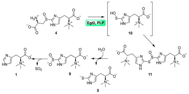
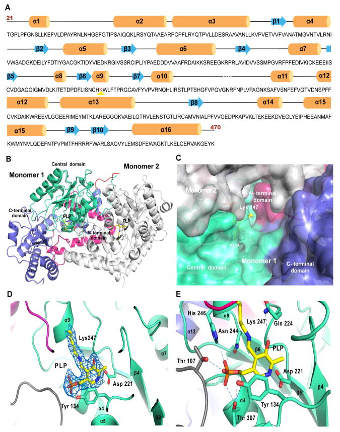
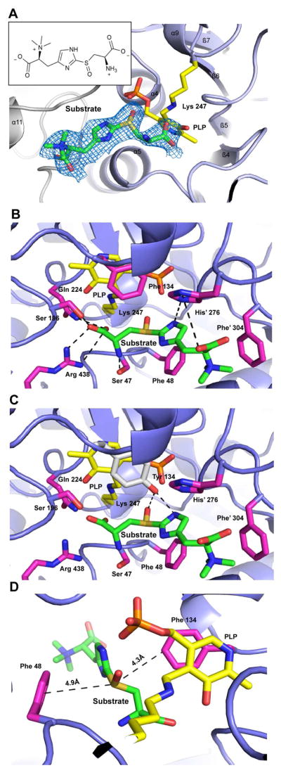
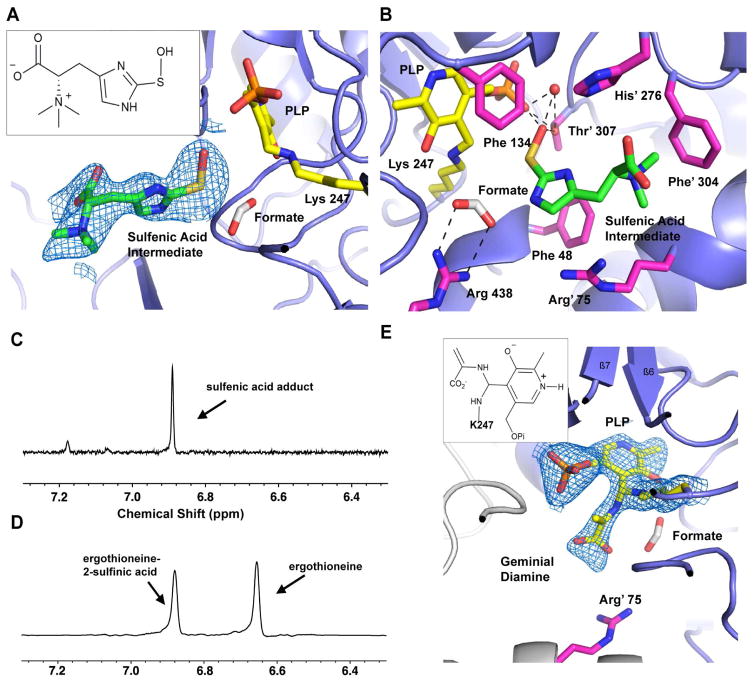
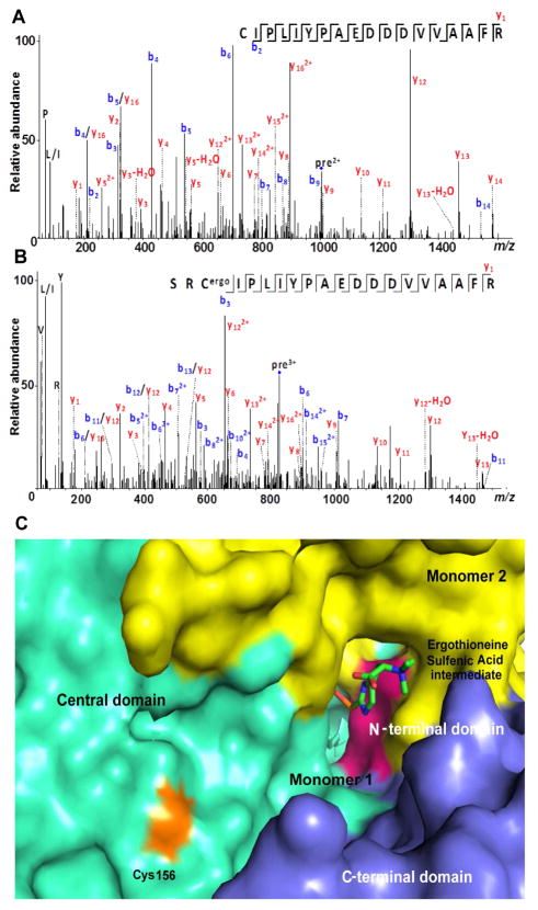
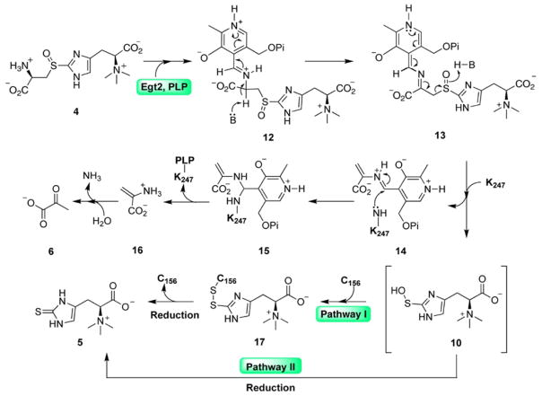
Sulfur incorporation in the biosynthesis of ergothioneine, a histidine thiol derivative, differs from other well-characterized transsulfurations. A combination of a mononuclear non-heme iron enzyme-catalyzed oxidative C-S bond formation and a subsequent pyridoxal 5'-phosphate (PLP)-mediated C-S lyase reaction leads to the net transfer of a sulfur atom from a cysteine to a histidine. In this study, we structurally and mechanistically characterized a PLP-dependent C-S lyase Egt2, which mediates the sulfoxide C-S bond cleavage in ergothioneine biosynthesis. A cation-π interaction between substrate and enzyme accounts for Egt2's preference of sulfoxide over thioether as a substrate. Using mutagenesis and structural biology, we captured three distinct states of the Egt2 C-S lyase reaction cycle, including a labile sulfenic intermediate captured in Egt2 crystals. Chemical trapping and high-resolution mass spectrometry were used to confirm the involvement of the sulfenic acid intermediate in Egt2 catalysis.

Keywords: C-S bond cleavage; X-ray crystallography; biosynthetic pathway; chemical trapping; enzymology; ergothioneine.


Conflict of interest statement

Declaration of Interests-

The authors declare competing financial interests. A patent (WO 2014100752 A1) on ergothioneine production through metabolic engineering has been submitted.

Figures

Fig. 1
Fig. 1
The proposed mechanistic model for Egt2 reaction using sulfoxide 4 as the substrate in the absence of reductant. The ergothioneine sulfenic acid intermediate is in brackets.
Fig. 2
Fig. 2. Overall Egt2 structure
A, The secondary structural elements of Egt2 are mapped along its sequence. α helices are represented by orange cylinders, β sheets by blue arrows, interconnecting loops as black lines and unstructured regions as black dashed lines. The PLP binding lysine is highlighted by red-color. The first 20 residues (residue 1–20) and the last 3 residues (residue 471–473) have not been shown since they were not visible in the crystal structure. B, A ribbon representation of the overall structure of the Egt2 dimer. For Monomer 1 the N-terminal domain (residue 21–99) is shown in pink, the central domain (residue 100–282) in green, the C-terminal domain (residue 293–470) in blue and the inter-domain loop (residue 283–294) is shown in red. The secondary structure elements are labeled and numbered. The cofactor PLP and K247 adduct are represented as yellow sticks. Monomer 2 is shown in grey. C, Surface representation of the active site of Egt2 in monomer 1. K247 and PLP are represented as yellow sticks. The color scheme is the same as the same as that in 2B. D, Internal aldimine formed by K247 and PLP are shown with 2mFo-DFc map contoured to 1σ. The PLP interacting residues D221 and the Y134 are shown in sticks. The color scheme is the same as that in 2B. E, The Egt2 cofactor PLP interaction network. Residues interacting with PLP were represented as sticks and hydrogen bonds with PLP are shown as blue dash lines. The color scheme is the same as that in 2B.
Fig. 3
Fig. 3. Structure of the Egt2 Y134F •substrate 4 binary complex
A, Sulfoxide substrate 4 in Egt2 Y134 active site shown with 2mFo-DFc map contoured to 0.75σ. The PLP cofactor is shown as yellow sticks. Monomer 2 is colored grey. Inset is the 2D chemical structure of the substrate 4. B, Interactions between Egt2 Y134 and substrate 4. Residues interacting with the substrate are shown as sticks in pink and residues from the neighboring monomer are indicated with a prime label. Hydrogen bonds between residues are shown by black dash lines. PLP and the K247 are shown as sticks in yellow. C, Interactions between Egt2 Y134F with tyrosine modeled at F134 position (shown as grey sticks) and its substrate 4. Residues interacting with the substrate are shown in pink and residues from the neighboring monomer are indicated with a prime label. Hydrogen bonds between tyrosine at the 134 position and the substrate are shown by black dash lines. PLP and the K247 are shown as yellow sticks. D, Cation-π interactions between the substrate and the aromatic side chains of F48 and F134. Distances between the cation and these π system are labeled.
Fig. 4
Fig. 4. Structure of the Egt2 Y134 in complex with a sulfenic acid intermediate
A, The ergothioneine sulfenic acid intermediate bound at the active site is shown with 2mFo-DFc map contoured to 0.8σ. The PLP is shown as yellow sticks. A formate ion is shown in white. Inset is the 2D chemical structure of the sulfenic acid intermediate 10. B, The interaction network of the ergothioneine sulfenic acid intermediate at the active site of Egt2 Y134F. Residues interacting with the substrate are shown as pink sticks. The formate ions are shown as grey sticks. Hydrogen bonds are shown by black dashed lines. PLP and the K247 are shown as yellow sticks. A water molecule hydrogen bonded to PLP and the sulfenic acid are shown as a red sphere. C – D, Egt2 reaction in the presence of 1,3-cyclohexanedione shows an appearance of a new peak at 6.86 ppm (C) while the control reaction without trapping reagent in D shows two signals at 6.65 ppm and 6.87, which correspond to ergothioneine 5 and ergothioneine-2-sulfinic acid 9 respectively. E, The 2mFo-DFc map of the geminial diamine intermediate contoured to 1σ. Inset is the 2D chemical structure of the geminial diamine intermediate 15.
Fig. 5
Fig. 5. Reactions of sulfenic acid intermediate
A, MS/MS spectrum of a tryptic peptide of the wild-type Egt2 (residue 156–173) before single turnover reaction. The parent ion has the signal with m/z 1003.998. B, MS/MS spectrum of a tryptic peptide of the wild-type Egt2 (residue 154–173) after the single turnover reaction. The non-specific cleavage of trypsin at S154 could be due to the presence of modification at C156. The parent ion has the signal with m/z 827.0616. C, Surface representation of the active site of Egt2 Y134 with the sulfenic acid intermediate shown in sticks and the C156 lying at the exit of the substrate binding pocket (colored in orange). Color scheme is identical to that in Fig. 2B and having C156 in orange.
Fig. 6
Fig. 6
The proposed Egt2 catalytic mechanism showing the involvement of the ergothioneine sulfenic acid, which could form a covalent adduct with C156 (Pathway I) or get released directly into solution (Pathway II). In both pathways, cellular thiol reduction system can reduce the intermediate to give ergothioneine as the final product. The ergothioneine sulfenic acid intermediate is in brackets.

References

    1. Allegrini A, Astegno A, La Verde V, Dominici P. Characterization of C-S lyase from Lactobacillus delbrueckii subsp bulgaricus ATCC BAA-365 and its potential role in food flavour applications. J Biochem. 2017;161:349–360. - PubMed
    1. Allison WS. Formation and reactions of sulfenic acids in proteins. Acc Chem Res. 1976;9:293–299.
    1. Aruoma OI, Spencer JPE, Mahmood N. Protection Against Oxidative Damage and Cell Death by the Natural Antioxidant Ergothioneine. Food Chem Toxicol. 1999;37:1043–1053. - PubMed
    1. Baldwin JE, Bradley M. Isopenicillin N synthase: mechanistic studies. Chem Rev. 1990;90:1079–1088.
    1. Bartholomeus Kuettner E, Hilgenfeld R, Weiss MS. Purification, characterization, and crystallization of alliinase from garlic. Arch Biochem Biophys. 2002;402:192–200. - PubMed

Publication types