. 2019 Feb;16(2):165-177.
doi: 10.1038/cmi.2017.167. Epub 2018 Mar 5.

Lymphatic endothelial cells regulate B-cell homing to lymph nodes via a NIK-dependent mechanism

Affiliations

Lymphatic endothelial cells regulate B-cell homing to lymph nodes via a NIK-dependent mechanism

Jie Yang et al. Cell Mol Immunol. 2019 Feb.

Abstract

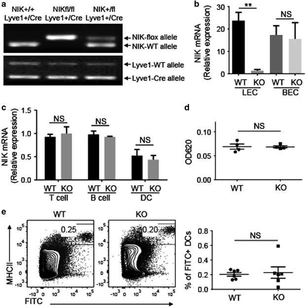
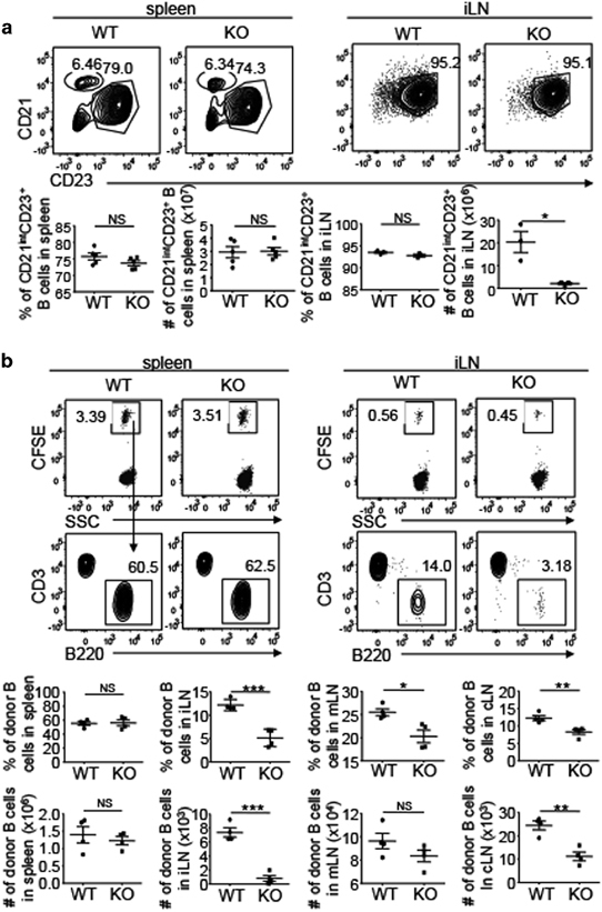
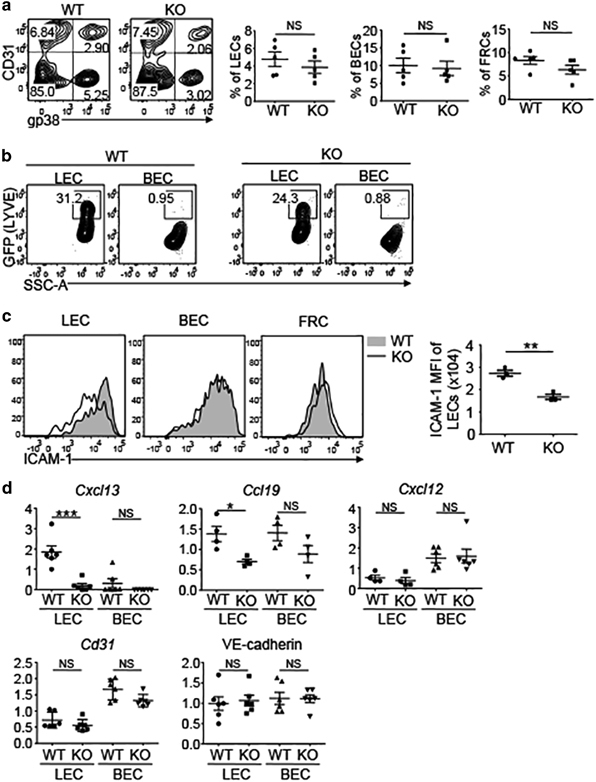
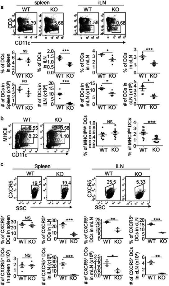
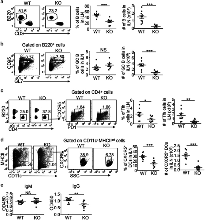
B cells home to the lymph nodes (LNs) via high endothelial venules (HEVs) under the guidance of chemokines, particularly CXCL13. However, as CXCL13 is not directly made in HEVs, the molecular mechanism mediating B-cell homing to LNs has remained unclear. We show here that nuclear factor (NF)-κB-inducing kinase (NIK), a kinase mediating activation of the noncanonical NF-κB pathway, functions in lymphatic endothelial cells (LECs) to regulate B-cell homing to LNs. LEC-conditional deletion of NIK in mice did not affect the integrity or global function of lymphatic vessels but caused a severe reduction in the frequency of B cells in LNs. The LEC-specific NIK deficiency did not affect the survival of B cells or the frequency of B cells in the spleen. B-cell adoptive transfer studies revealed that the LEC-specific NIK deletion impairs the ability of LNs to recruit B cells. We further show that NIK mediates expression of the chemokines CXCL13 and CCL19 in LECs. Although CCL19 is also expressed in blood endothelial cells (BECs), CXCL13 is not produced in BECs. These results suggest that NIK regulates naive B-cell homing to LNs via mediating production of the B-cell homing chemokine CXCL13 in LECs.


Conflict of interest statement

The authors declare no conflict of interest.

Figures

Figure 1
Figure 1
LEC-specific NIK is dispensable for global function of lymphatic vessels. (a) Genotyping PCR analysis of NIK-flox, NIK-WT, Lyve1-Cre and Lyve1-WT alleles using tail DNA to identify wild-type (WT, NIK+/+Lyve1+/Cre), heterozygous (NIK+/flLyve1+/Cre) and homozygous LEC-conditional NIK-KO (NIKLEC-KO, NIKfl/flLyve1+/Cre) mice. (b, c) The qRT-PCR analysis of Nik expression was performed using total RNA from LECs and BECs (b) or T cells, B cells and DCs (c) sorted from WT and NIKLEC-KO (KO) mice by flow cytometry. Data are presented as summary graphs for the expression of Nik normalized to the expression of the housekeeping gene Actb. (d) Measurement of lymph flow in the ear with Evan blue dye. Evan blue dye was injected to the ears of WT and NIKLEC-KO (KO) mice and the remaining dye was extracted 24 h after injection. The amount of extracted dye was determined by measuring the absorbance at 620 nm. (e) Flow cytometry analysis of the frequency of skin-derived DCs (FITC+MHCII+) in iLNs from WT and NIKLEC-KO (KO) mice 18 h after FITC painting. Data are presented as representative plots (left) and summary graphs (right). Data are representative of two independent experiments and are presented as means±s.e.m. BEC, blood endothelial cell; DC, dendritic cell; FITC, fluorescein isothiocyanate; iLN, inguinal lymph node; KO, knockout; LEC, lymphatic endothelial cell; Lyve1, lymphatic endothelial hyaluronan receptor 1; NIK, nuclear factor-κB-inducing kinase; NS, not significant, qRT-PCR, real-time quantitative PCR; WT, wild type. **P<0.01.
Figure 2
Figure 2
LEC-specific NIK deletion reduces the number of B cells in LNs. (a, b) Representative images (a) and total cell number (b) of spleen, iLNs, mLNs and cLNs of WT and NIKLEC-KO (KO) mice. (c) Flow cytometry analysis of the frequency and absolute cell number of T cells (CD3+) and B cells (B220+) in the spleen, iLN, mLN and cLN of WT and NIKLEC-KO (KO) mice. Data are presented as representative plots (upper panel) and summary graphs (lower panel). Data are representative of at least three independent experiments and are presented as means±s.e.m. *P<0.05; **P<0.01; ***P<0.001. cLN, cervical lymph node; DC, dendritic cell; iLN, inguinal lymph node; KO, knockout; LEC, lymphatic endothelial cell; LN, lymph node; mLN, mesenteric lymph node; NIK, nuclear factor-κB-inducing kinase; NS, not significant, WT, wild type.
Figure 3
Figure 3
LEC-specific NIK regulates B-cell homing to LNs. (a) Flow cytometry analysis marginal zone (CD21highCD23low) and follicular (CD21intCD23+) B cells in the spleen and follicular B cells in iLNs of WT (NIK+/+Lyve1+/Cre) and NIKLEC-KO (KO, NIKfl/flLyve1+/Cre) mice. Plots were gated on B220+ B cells. Summary graphs of the frequency and absolute cell number of follicular B cells in the spleen and iLNs are shown in the lower panels (each symbol represents a mouse). (b) Flow cytometry analysis of the frequency and absolute cell number of CFSE+ donor B cells (B220+) migrating to the spleen, iLN, mLN and cLN of WT and NIKLEC-KO (KO) recipient mice 4 h after adoptive transfer with CFSE-labeled WT splenocytes. Data are presented as representative plots (upper panel) and summary graphs (lower panel). Data are representative of two independent experiments and are presented as means±s.e.m. *P<0.05; **P<0.01; ***P<0.001. CFSE, carboxyfluorescein succinimidyl ester; cLN, cervical lymph node; DC, dendritic cell; iLN, inguinal lymph node; KO, knockout; LEC, lymphatic endothelial cell; LN, lymph node; mLN, mesenteric lymph node; NIK, nuclear factor-κB-inducing kinase; NS, not significant, WT, wild type.
Figure 4
Figure 4
NIK regulates the expression of CXCL13 in LECs. (a) Flow cytometry analysis of the frequency of LECs (CD31+gp38+), BECs (CD31+gp38) and FRCs (CD31-gp38+) in the LNs of WT and NIKLEC-KO (KO) mice. Plots were gated on CD45.2 stromal cells. Data are presented as representative plots (left) and summary graphs (right). (b) Flow cytometry analysis of the expression of Lyve1 (represented by GFP expression) by LECs and BECs in the LNs of WT and NIKLEC-KO (KO) mice. (c) Flow cytometry analysis of the expression of ICAM-1 by LECs, BECs and FRCs in the LNs of WT and NIKLEC-KO (KO) mice. Data are presented as representative plots (left) and summary graphs for the MFI of ICAM-1 (right). (d) qRT-PCR analysis was performed using total RNA from LECs and BECs that were sorted from LNs of WT and NIKLEC-KO (KO) mice by flow cytometry. Data are presented as summary graphs for the expression of indicated genes normalized to the expression of the housekeeping gene Actb. Data are representative of at least three independent experiments and are presented as means±s.e.m. *P<0.05; **P<0.01; ***P<0.001. BEC, blood endothelial cell; FRC, fibroblast reticular cell; ICAM-1, intercellular adhesion molecule 1; LN, lymph node; KO, knockout; LEC, lymphatic endothelial cell; Lyve1, lymphatic endothelial hyaluronan receptor 1; MFI, mean fluorescence intensity; NIK, nuclear factor-κB-inducing kinase; NS, not significant, qRT-PCR, real-time quantitative PCR; WT, wild type.
Figure 5
Figure 5
NIK deletion in LECs impairs the homing of CXCR5+ DCs. (a) Flow cytometry analysis of the frequency and absolute cell number of DCs (CD3-CD11c+) in the spleen, iLN, mLN and cLN of WT and NIKLEC-KO (KO) mice. Data are presented as representative plots (upper panel) and summary graphs (lower panel). (b) Flow cytometry analysis of the frequency of MHCIIhighCD11c+ and MHCIIintCD11c+ DCs in the iLN of WT and NIKLEC-KO (KO) mice. Data are presented as representative plots (left) and summary graphs (right). (c) Flow cytometry analysis of the frequency and absolute cell number of CXCR5+ DCs in the spleen, iLN, mLN and cLN of WT and NIKLEC-KO (KO) mice. Plots are gated on MHCIIintCD11c+ DCs. Data are presented as representative plots (upper panel) and summary graphs (lower panel). Data are representative of at least three independent experiments and are presented as means±s.e.m. *P<0.05; **P<0.01; ***P<0.001. cLN, cervical lymph node; DC, dendritic cell; iLN, inguinal lymph node; KO, knockout; LEC, lymphatic endothelial cell; mLN, mesenteric lymph node; NIK, nuclear factor-κB-inducing kinase; NS, not significant.
Figure 6
Figure 6
Effect of LEC-specific NIK deficiency on immune responses. (a) Flow cytometry analysis of the frequency and absolute cell number of B cells (B220+) in the iLNs of WT and NIKLEC-KO (KO) mice immunized with NP-KLH. Data are presented as representative plots (left) and summary graphs (right). (b–d) Flow cytometry analysis of the frequency and absolute cell number of germinal center B cells (b, CD95+GL7+), TFH cells (c, PD1+CXCR5+) and CXCR5+ DCs (d) in the iLNs of WT and NIKLEC-KO (KO) mice immunized with NP-KLH. Plots were gated on B220+ (b), CD4+ (c) and CD11c+MHCIIint (d) cells. Data are presented as representative plots (left) and summary graphs (right). (e) ELISA of NP-specific IgM and IgG in the sera of WT and NIKLEC-KO (KO) mice immunized with NP-KLH. Data are representative of two independent experiments and are presented as means±s.e.m. *P<0.05; **P<0.01; ***P<0.001. DC, dendritic cell; ELISA, enzyme-linked immunosorbent assay; Ig, immunoglobulin; iLN, inguinal lymph node; KO, knockout; LEC, lymphatic endothelial cell; NIK, nuclear factor-κB-inducing kinase; NP-KLH, hapten NP (4-hydroxy-3-nitrophenylacetyl) linked to keyhole lympet hemocyanin; NS, not significant, TFH, T follicular helper; WT, wild type.

References

    1. Gowans JL. The recirculation of lymphocytes from blood to lymph in the rat. J Physiol. 1959;146:54–69. doi: 10.1113/jphysiol.1959.sp006177. - DOI - PMC - PubMed
    1. Girard JP, Springer TA. High endothelial venules (HEVs): specialized endothelium for lymphocyte migration. Immunol Today. 1995;16:449–457. doi: 10.1016/0167-5699(95)80023-9. - DOI - PubMed
    1. Miyasaka M, Tanaka T. Lymphocyte trafficking across high endothelial venules: dogmas and enigmas. Nat Rev Immunol. 2004;4:360–370. doi: 10.1038/nri1354. - DOI - PubMed
    1. Girard JP, Moussion C, Forster R. HEVs, lymphatics and homeostatic immune cell trafficking in lymph nodes. Nat Rev Immunol. 2012;12:762–773. doi: 10.1038/nri3298. - DOI - PubMed
    1. von Andrian UH, Mempel TR. Homing and cellular traffic in lymph nodes. Nat Rev Immunol. 2003;3:867–878. doi: 10.1038/nri1222. - DOI - PubMed

Publication types

MeSH terms

LinkOut - more resources