

Culturable mycobiota from Karst caves in China, with descriptions of 20 new species
- PMID: 29503468
- PMCID: PMC5832949
- DOI: 10.3767/persoonia.2017.39.01
Culturable mycobiota from Karst caves in China, with descriptions of 20 new species
Abstract
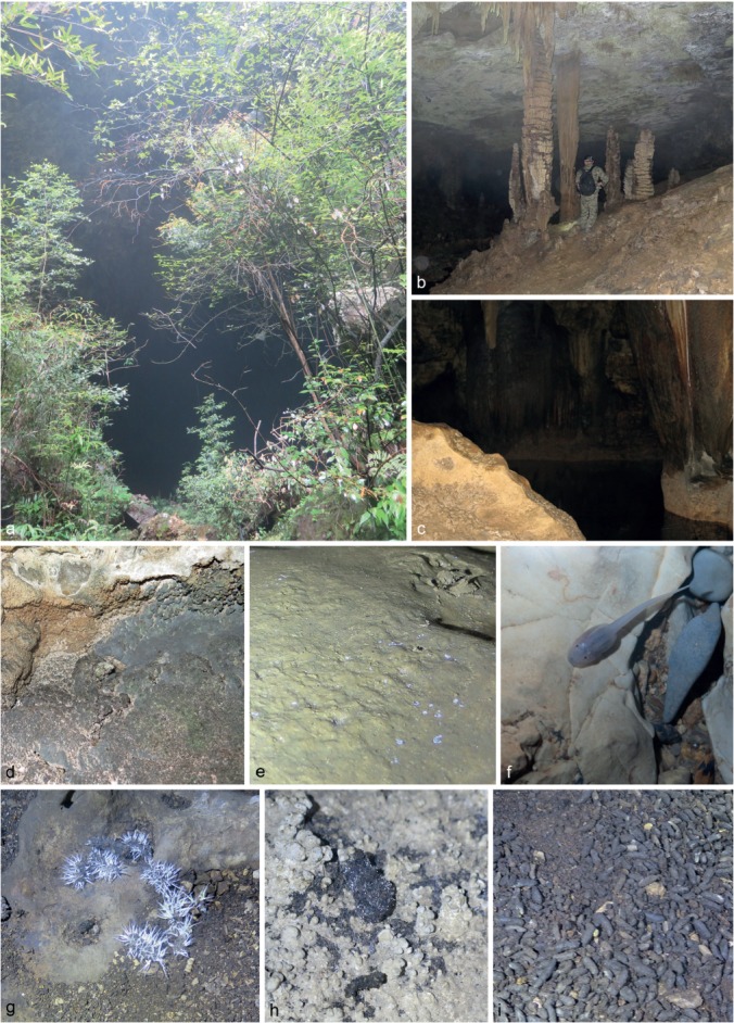
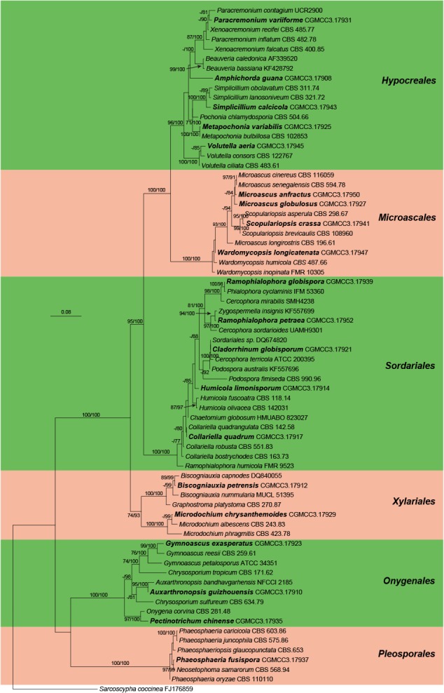
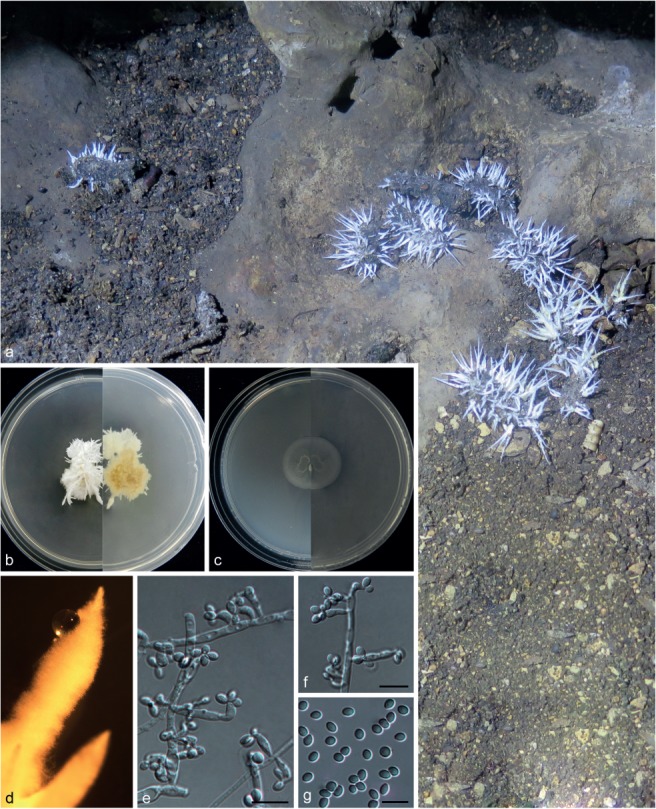
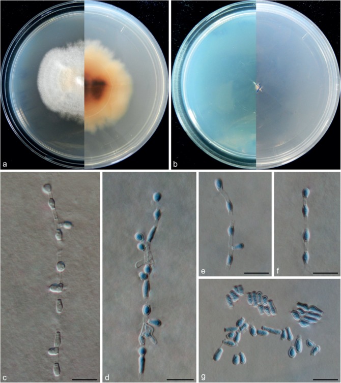
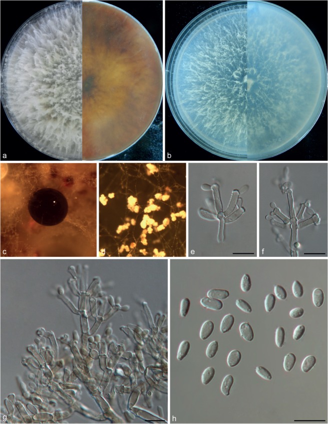
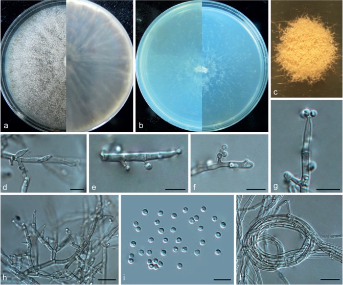
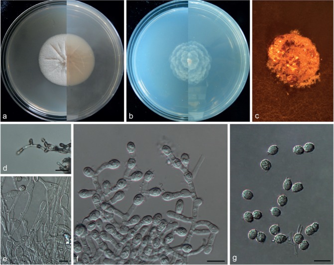
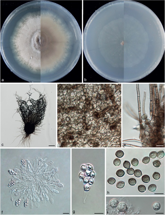
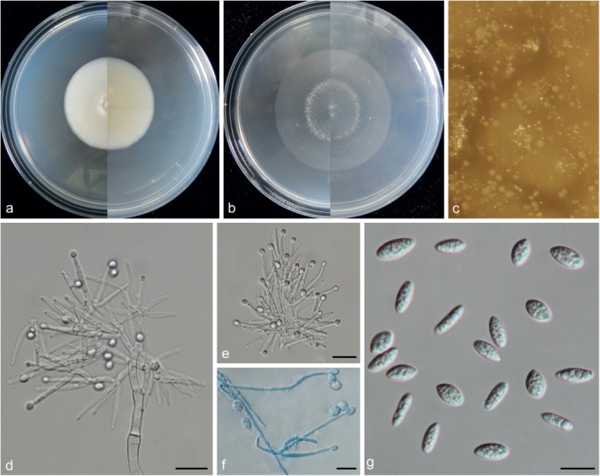
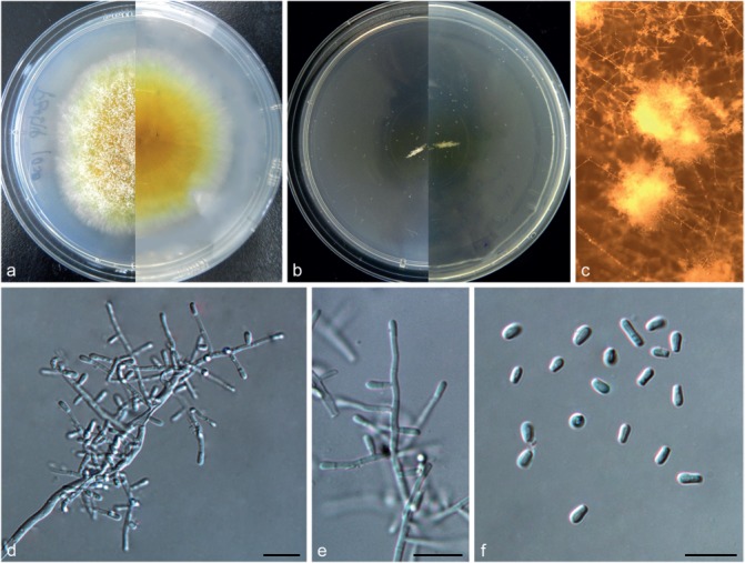
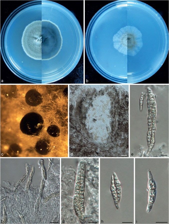
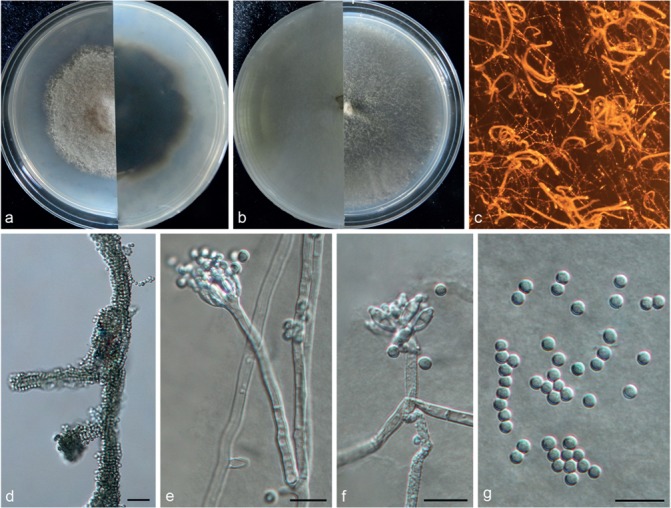
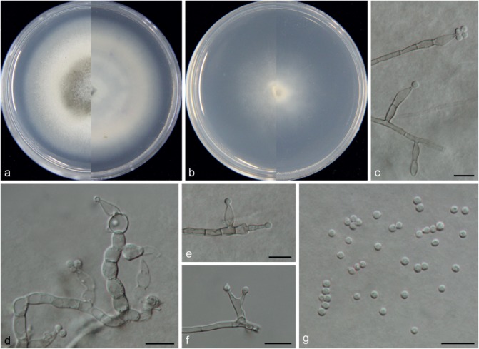
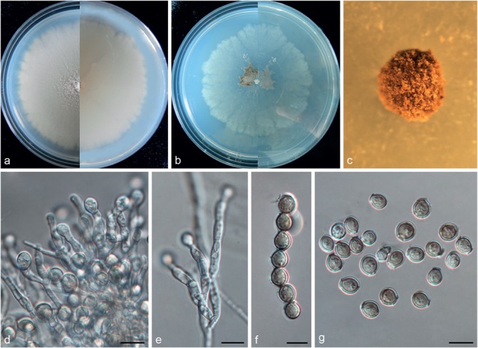
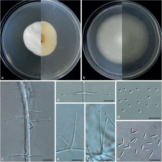
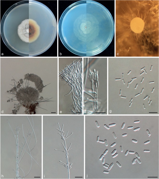
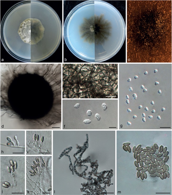
Karst caves are distinctly characterised by darkness, low to moderate temperatures, high humidity, and scarcity of organic matter. During the years of 2014-2015, we explored the mycobiota in two unnamed Karst caves in Guizhou province, China, and obtained 563 fungal strains via the dilution plate method. Preliminary ITS analyses of these strains suggested that they belonged to 246 species in 116 genera, while 23.5 % were not identified to species level. Among these species, 85.8 % (211 species) belonged to Ascomycota; 7.3 % (18 species) belonged to Basidiomycota; 6.9 % (17 species) belonged to Mucoromycotina. The majority of these species have been previously known from other environments, mostly from plants or animals as pathogens, endophytes or via a mycorrhizal association. We also found that 59 % of these species were discovered for the first time from Karst caves, including 20 new species that are described in this paper. The phylogenetic tree based on LSU sequences revealed 20 new species were distributed in six different orders. In addition, ITS or multi-locus sequences were employed to infer the phylogenetic relationships of new taxa with closely related allies. We conclude that Karst caves encompass a high fungal diversity, including a number of previously unknown species. Novel species described include: Amphichorda guana, Auxarthronopsis guizhouensis, Biscogniauxia petrensis, Cladorrhinum globisporum, Collariella quadrum, Gymnoascus exasperatus, Humicola limonisporum, Metapochonia variabilis, Microascus anfractus, Microascus globulosus, Microdochium chrysanthemoides, Paracremonium variiforme, Pectinotrichum chinense, Phaeosphaeria fusispora, Ramophialophora globispora, Ramophialophora petraea, Scopulariopsis crassa, Simplicillium calcicola, Volutella aeria, and Wardomycopsis longicatenata.
Keywords: ITS DNA barcodes; diversity; morphology; systematics; troglobitic fungi.
Figures

Similar articles
-
Origin of Cave Fungi.Front Microbiol. 2018 Jun 28;9:1407. doi: 10.3389/fmicb.2018.01407. eCollection 2018. Front Microbiol. 2018. PMID: 30013527 Free PMC article.
-
Eight novel cave fungi in Thailand's Satun Geopark.Fungal Syst Evol. 2023 Nov;12:1-30. doi: 10.3114/fuse.2023.12.01. Epub 2023 Jul 24. Fungal Syst Evol. 2023. PMID: 38455950 Free PMC article.
-
Unravelling the hidden diversity of cave mycobiota in Thailand's Satun Geopark.Sci Rep. 2023 Nov 6;13(1):19162. doi: 10.1038/s41598-023-43316-2. Sci Rep. 2023. PMID: 37932293 Free PMC article.
-
Unveiling the Subterranean Symphony: A Comprehensive Study of Cave Fungi Revealed Through National Center for Biotechnology Sequences.J Fungi (Basel). 2025 Apr 5;11(4):286. doi: 10.3390/jof11040286. J Fungi (Basel). 2025. PMID: 40278107 Free PMC article. Review.
-
A new species of Tachycines Adelung, 1902 (Orthoptera, Rhaphidophoridae, Aemodogryllinae, Aemodogryllini) from karst caves in Guizhou, China.Zookeys. 2020 Jun 1;937:21-29. doi: 10.3897/zookeys.937.49173. eCollection 2020. Zookeys. 2020. PMID: 32547297 Free PMC article. Review.
Cited by
-
New and Interesting Fungi. 4.Fungal Syst Evol. 2021 Jun;7:255-343. doi: 10.3114/fuse.2021.07.13. Epub 2021 Apr 28. Fungal Syst Evol. 2021. PMID: 34124627 Free PMC article.
-
Origin of Cave Fungi.Front Microbiol. 2018 Jun 28;9:1407. doi: 10.3389/fmicb.2018.01407. eCollection 2018. Front Microbiol. 2018. PMID: 30013527 Free PMC article.
-
Three New Species of Microdochium (Sordariomycetes, Amphisphaeriales) on Miscanthus sinensis and Phragmites australis from Hainan, China.J Fungi (Basel). 2022 May 27;8(6):577. doi: 10.3390/jof8060577. J Fungi (Basel). 2022. PMID: 35736060 Free PMC article.
-
Phylogenetic diversity and morphological characterization of cordycipitaceous species in Taiwan.Stud Mycol. 2024 Dec;109:1-56. doi: 10.3114/sim.2024.109.01. Epub 2024 Jun 19. Stud Mycol. 2024. PMID: 39717658 Free PMC article.
-
Two new species of Microdochium from Indocalamus longiauritus in south-western China.MycoKeys. 2020 Sep 10;72:93-108. doi: 10.3897/mycokeys.72.55445. eCollection 2020. MycoKeys. 2020. PMID: 32982557 Free PMC article.
References
-
- Ajello L, Briceño-Maaz T, Campins H, et al. 1960a. Isolation of Histoplasma capsulatum from an oil bird (Steatornis caripensis) cave in Venezuela. Mycopathologia et Mycologia Applicata 12: 199–206. - PubMed
-
- Ajello L, Manson-Bahr PEC, Moore JC. 1960b. Amboni caves, Tanganyika, a new endemic area for Histoplasma capsulatum. American Journal of Tropical Medicine and Hygiene 9: 633–638. - PubMed
-
- Al-Doory Y, Rhoades ER. 1968. Isolation of Histoplasma capsulatum from a Texas cave. Mycopathologia et Mycologia Applicata 35: 201–207. - PubMed
-
- Bai G, Shaner G. 2004. Management and resistance in wheat and barley to Fusarium head blight. Annual Review of Phytopathology 42: 135–161. - PubMed
-
- Barton HA, Jurado V. 2007. What’s up down there? Microbial diversity in caves. Microbe 2: 132–138.
LinkOut - more resources
Full Text Sources
Other Literature Sources
Molecular Biology Databases