. 2018 Feb 16:9:243.
doi: 10.3389/fimmu.2018.00243. eCollection 2018.

The Rac Activator DOCK2 Mediates Plasma Cell Differentiation and IgG Antibody Production

Affiliations

The Rac Activator DOCK2 Mediates Plasma Cell Differentiation and IgG Antibody Production

Miho Ushijima et al. Front Immunol. .

Abstract

A hallmark of humoral immune responses is the production of antibodies. This process involves a complex cascade of molecular and cellular interactions, including recognition of specific antigen by the B cell receptor (BCR), which triggers activation of B cells and differentiation into plasma cells (PCs). Although activation of the small GTPase Rac has been implicated in BCR-mediated antigen recognition, its precise role in humoral immunity and the upstream regulator remain elusive. DOCK2 is a Rac-specific guanine nucleotide exchange factor predominantly expressed in hematopoietic cells. We found that BCR-mediated Rac activation was almost completely lost in DOCK2-deficient B cells, resulting in defects in B cell spreading over the target cell-membrane and sustained growth of BCR microclusters at the interface. When wild-type B cells were stimulated in vitro with anti-IgM F(ab')2 antibody in the presence of IL-4 and IL-5, they differentiated efficiently into PCs. However, BCR-mediated PC differentiation was severely impaired in the case of DOCK2-deficient B cells. Similar results were obtained in vivo when DOCK2-deficient B cells expressing a defined BCR specificity were adoptively transferred into mice and challenged with the cognate antigen. In addition, by generating the conditional knockout mice, we found that DOCK2 expression in B-cell lineage is required to mount antigen-specific IgG antibody. These results highlight important role of the DOCK2-Rac axis in PC differentiation and IgG antibody responses.

Keywords: B cell receptor; DOCK2; Rac activation; antibody production; immunological synapse; plasma cell.


Figures

Figure 1
Figure 1
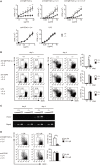
DOCK2 is a major Rac GEF acting downstream of B cell receptor (BCR). (A) BCR-mediated activation of Rac1 and Rac2 were compared between Dock2+/+ and Dock2−/− lymph node (LN) B cells. Results were quantified by densitometry and are expressed as the ratio of GTP-bound form to the total protein after normalization of the 0.5 min-value of Dock2+/+ samples to an arbitrary value of 1. Data for 0.5 min are indicated as the mean ± SD of five independent experiments. **p < 0.01 (two-tailed Mann–Whitney test). (B) Fura-2-loaded Dock2+/+ and Dock2−/− LN B cells were stimulated with anti-IgM F(ab′)2 antibody or ionomycin. Data are indicated as the Fura-2 ratio at 340:380 nm and are representative of three independent experiments. (C,D) Dock2+/+ and Dock2−/− LN B cells were stimulated with anti-IgM F(ab′)2 antibody and analyzed for phosphorylation of each molecule. In (C), phosphorylations of ERK, Syk, Akt, BLNK, and CD19 were analyzed using phosphorylation-specific antibodies. In (D), cell extracts were immunoprecipitated with anti-Vav or anti-PLCγ2 antibody and analyzed with anti-phosphotyrosine antibody.
Figure 2
Figure 2
DOCK2 regulates B cell receptor-mediated B cell proliferation and PC differentiation in vitro. (A) Dock2+/+ and Dock2−/− LN B cells were stimulated with anti-IgM F(ab′)2 antibody, anti-CD40 antibody, or lipopolysaccharide at the indicated concentrations in the presence or absence of IL-4/IL-5, and B cell proliferation was analyzed. Data are indicated as the mean ± SD of five independent experiments. *p < 0.05; **p < 0.01 (two-tailed Mann–Whitney test). (B) Following stimulation with anti-IgM F(ab′)2 or anti-CD40 antibody in the presence of IL-4 and IL-5 for 4 days, Dock2+/+ and Dock2−/− LN B cells were analyzed for the expression of CD138 and B220 to assess PC differentiation. FACS profiles at day 0 and day 4 after stimulation are shown. Data are indicated as the mean ± SD of five independent experiments. **p < 0.01 (two-tailed unpaired Student’s t-test). (C) Following stimulation of Dock2+/+ and Dock2−/− LN B cells with anti-IgM F(ab′)2 antibody in the presence of IL-4 and IL-5, the expression of Prdm1 and Gapdh were analyzed with reverse transcription-PCR. Amplification increased by three cycles from the left to the right starting at 28 cycles for Prdm1 or 20 cycles for Gapdh. Data are representative of three independent experiments. (D) The effect of CPYPP (12.5 µM) on in vitro PC differentiation was analyzed as in (B). Data are indicated as the mean ± SD of seven independent experiments. **p < 0.01 (two-tailed Mann–Whitney test).
Figure 3
Figure 3
DOCK2 regulates immunological synapse formation. (A) FACS profiles showing comparable binding of HEL to Dock2+/− and Dock2−/− LN B cells. LN B cells from C57BL/6 mice were used as control samples. (B–D) Following incubation of Dock2+/− and Dock2−/− HyHEL10 mice with baby hamster kidney (BHK)-ICAM-HEL, the area of B cell contact (C) and the mean fluorescence intensity (MFI) of HEL-GFP (D) were compared at the indicated time points. Data are indicated as the mean ± SD of 60 cells collected from three separate experiments. **p < 0.01 (two-tailed Mann–Whitney test). (E,F) Following incubation of Dock2+/− and Dock2−/− HyHEL10 mice with BHK-ICAM-HEL, the MFI of leukocyte function-associated antigen-1 (LFA-1) (F) was compared at the indicated time points. Data are indicated as the mean ± SD of 60 cells collected from three separate experiments. *p < 0.05; **p < 0.01 (two-tailed Mann–Whitney test).
Figure 4
Figure 4
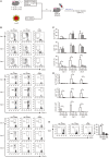
DOCK2 is required for germinal center (GC) B cell expansion and PC differentiation in adoptive transfer model. (A) Schematic representation of the adoptive transfer model used in this study. (B) FACS profiles indicating the expression of GL7, CD38, IgG1, and/or CD138 in CD45.1+ transferred B cells from Dock2+/− and Dock2−/− HyHEL10 mice. Data were obtained at day 4, day 5, and day 6 after transfer and are representative of five independent experiments. (C) Following adoptive transfer, the percentages of GL7+CD38 GC B cells, IgG1+ B cells, CD138+ plasma cells (PCs) in CD45.1+ transferred B cells were compared at the indicated time points between Dock2+/− and Dock2−/− HyHEL10 mice. Data are indicated as the mean ± SD of five independent experiments. *p < 0.05; **p < 0.01 (two-tailed Mann–Whitney test). (D) Following adoptive transfer, the numbers of GL7+CD38 GC B cells, IgG1+ B cells, CD138+ PCs were compared at the indicated time points between Dock2+/− and Dock2−/− HyHEL10 mice. Data are indicated as the mean ± SD of five independent experiments. **p < 0.01 (two-tailed Mann–Whitney test). (E) The percentages of BrdU+ B cells in CD45.1+ transferred B cells were compared between Dock2+/− and Dock2−/− HyHEL10 mice 5 days after adoptive transfer. Data are indicated as the mean ± SD of nine independent experiments. **p < 0.01 (two-tailed Mann–Whitney test).
Figure 5
Figure 5
Characterization of CD19-Cre+/− Dock2lox/lox mice. (A) FACS profiles for expression of IgM and IgD in the CD19+ bone marrow B cells. The number of each subset of B cells was compared between CD19-Cre−/− Dock2lox/lox and CD19-Cre+/− Dock2lox/lox mice. Data are indicated as the mean ± SD of five mice. **p < 0.01 (two-tailed Mann–Whitney test). (B) FACS profiles for expression of IgM and IgD in the B220+ splenic B cells. The number of each subset of B cells was compared between CD19-Cre−/− Dock2lox/lox and CD19-Cre+/− Dock2lox/lox mice. Data are indicated as the mean ± SD of 5 mice. **p < 0.01 (two-tailed unpaired Student’s t-test). (C) FACS profiles for expression of CD21 and heat stable antigen (HSA) in the B220+ splenic B cells. The number of each subset of B cells (T1, T2, and follicular B cells) was compared between CD19-Cre–/– Dock2lox/lox and CD19-Cre+/− Dock2lox/lox mice. Data are indicated as the mean ± SD of five mice. *p < 0.05; **p < 0.01 (two-tailed Mann–Whitney test). (D) FACS profiles for expression of IgM and IgD or CD21 and HSA in the B220+ peripheral LN (PLN) B cells. The number of each subset of B cells was compared between CD19-Cre−/− Dock2lox/lox and CD19-Cre+/− Dock2lox/lox mice. Data are indicated as the mean ± SD of five mice. *p < 0.05; **p < 0.01 (two-tailed Mann–Whitney test). (E) Immunohistochemical analyses of the spleen sections from CD19-Cre−/− Dock2lox/lox and CD19-Cre+/− Dock2lox/lox mice.
Figure 6
Figure 6
Defective antibody production in CD19-Cre+/− Dock2lox/lox mice. (A) Comparison of serum IgM, IgG1 and IgG2b in CD19-Cre−/− Dock2lox/lox and CD19-Cre+/− Dock2lox/lox mice under the steady state. Data are indicated as the mean ± SD of 10 mice. **p < 0.01 (two-tailed Mann–Whitney test). (B) OVA-specific antibody production was compared between CD19-Cre−/− Dock2lox/lox and CD19-Cre+/− Dock2lox/lox mice at day 14 after immunization. For negative controls, wells were coated with HEL. Data are indicated as the mean ± SD of five independent experiments. *p < 0.05; **p < 0.01 (two-tailed Mann–Whitney test).

Similar articles

Cited by

References

    1. Hardy RR, Carmack CE, Shinton SA, Kemp JD, Hayakawa K. Resolution and characterization of pro-B and pre-pro-B cell stages in normal mouse bone marrow. J Exp Med (1991) 173(5):1213–25.10.1084/jem.173.5.1213 - DOI - PMC - PubMed
    1. Loder F, Mutschler B, Ray RJ, Paige CJ, Sideras P, Torres R, et al. B cell development in the spleen takes place in discrete steps and is determined by the quality of B cell receptor-derived signals. J Exp Med (1999) 190(1):75–89.10.1084/jem.190.1.75 - DOI - PMC - PubMed
    1. Reth M, Wienands J. Initiation and processing of signals from the B cell antigen receptor. Annu Rev Immunol (1997) 15:453–79.10.1146/annurev.immunol.15.1.453 - DOI - PubMed
    1. Harwood NE, Batista FD. Early events in B cell activation. Annu Rev Immunol (2010) 28:185–210.10.1146/annurev-immunol-030409-101216 - DOI - PubMed
    1. MacLennan ICM, Toellner KM, Cunningham AF, Serre K, Sze DMY, Zúñiga E, et al. Extrafollicular antibody responses. Immunol Rev (2003) 194:8–18.10.1034/j.1600-065X.2003.00058.x - DOI - PubMed

Publication types

MeSH terms