Review
. 2018 Feb 19:16:Doc04.
doi: 10.3205/cto000143. eCollection 2017.

Cochlear implant - state of the art

Affiliations
Review

Cochlear implant - state of the art

Thomas Lenarz. GMS Curr Top Otorhinolaryngol Head Neck Surg. .

Abstract

Cochlear implants are the treatment of choice for auditory rehabilitation of patients with sensory deafness. They restore the missing function of inner hair cells by transforming the acoustic signal into electrical stimuli for activation of auditory nerve fibers. Due to the very fast technology development, cochlear implants provide open-set speech understanding in the majority of patients including the use of the telephone. Children can achieve a near to normal speech and language development provided their deafness is detected early after onset and implantation is performed quickly thereafter. The diagnostic procedure as well as the surgical technique have been standardized and can be adapted to the individual anatomical and physiological needs both in children and adults. Special cases such as cochlear obliteration might require special measures and re-implantation, which can be done in most cases in a straight forward way. Technology upgrades count for better performance. Future developments will focus on better electrode-nerve interfaces by improving electrode technology. An increased number of electrical contacts as well as the biological treatment with regeneration of the dendrites growing onto the electrode will increase the number of electrical channels. This will give room for improved speech coding strategies in order to create the bionic ear, i.e. to restore the process of natural hearing by means of technology. The robot-assisted surgery will allow for high precision surgery and reliable hearing preservation. Biological therapies will support the bionic ear. Methods are bio-hybrid electrodes, which are coded by stem cells transplanted into the inner ear to enhance auto-production of neurotrophins. Local drug delivery will focus on suppression of trauma reaction and local regeneration. Gene therapy by nanoparticles will hopefully lead to the preservation of residual hearing in patients being affected by genetic hearing loss. Overall the cochlear implant is a very powerful tool to rehabilitate patients with sensory deafness. More than 1 million of candidates in Germany today could benefit from this high technology auditory implant. Only 50,000 are implanted so far. In the future, the procedure can be done under local anesthesia, will be minimally invasive and straight forward. Hearing preservation will be routine.

Keywords: cochlear implant; complications; diagnostics; future developments; results; sensory deafness; surgical procedure.


Conflict of interest statement

The author declares that he has no competing interests.

Figures

Table 1
Table 1. Preoperative diagnostics for indication of cochlear implantation in adult (A) and pediatric (P) patients
Table 2
Table 2. Constellation of findings in cases of perisynaptic audiopathy
Table 3
Table 3. Malformations of the inner ear and the temporal bone and cochlear implantation based on Sennaroglu and Saatci, 2003 [42]
Table 4
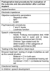
Table 4. Test procedures for assessment of hearing performance
Table 5
Table 5. Postoperative complications
Table 6
Table 6. Risks in children
Figure 1
Figure 1. Cochlear implant system, overview (courtesy of Cochlear Company)
Figure 2
Figure 2. Bottleneck of the electrode-nerve interface (according to A. Büchner)
Figure 3
Figure 3. Neural response telemetry (courtesy of Cochlear Company)
Figure 4
Figure 4. CI electrodes of different lengths
Figure 5
Figure 5. Peri-modiolar electrode
Figure 6
Figure 6. Technical realization of remote fitting (according to A. Büchner). Expert center of the Hannover Medical School. Decentral hearing center, based on the Auric system, coded data connection, secured bandwidth (2 Mbit SDSL)
Figure 7
Figure 7. Cone beam tomography of the temporal bone
Figure 8
Figure 8. MRI of the temporal bone, T2 image of the cochlea and the internal auditory meatus
Figure 9
Figure 9. MRI, T2 image of a transversal section through the internal auditory meatus. Aplasia of the hearing nerve on the right side (right image) and normal findings on the left with acoustic nerve (left below), facial nerve (left above), and vestibular nerve (right)
Figure 10
Figure 10. Retroauricular incision. Periostal flap. Periostal pouch.
Figure 11
Figure 11. Mastoidectomy with cortical projection (courtesy of Endo-Press, Tuttlingen, Germany)
Figure 12
Figure 12. Creation of the bone bed (courtesy of Endo-Press, Tuttlingen, Germany)
Figure 13
Figure 13. Creation of a connecting tunnel/canal from the bone bed to the mastoid (courtesy of Endo-Press, Tuttlingen, Germany)
Figure 14
Figure 14. Posterior tympanostomy (courtesy of Endo-Press, Tuttlingen, Germany)
Figure 15
Figure 15. Insertion of the implant (courtesy of Endo-Press, Tuttlingen, Germany)
Figure 16
Figure 16. Exposition of the round window membrane (courtesy of Endo-Press, Tuttlingen, Germany)
Figure 17
Figure 17. Cochleostomy (courtesy of Endo-Press, Tuttlingen, Germany)
Figure 18
Figure 18. Insertion of the electrode through the round window
Figure 19
Figure 19. Electrode insertion by so-called advanced-off stylet technique (courtesy of Endo-Press, Tuttlingen, Germany)
Figure 20
Figure 20. Fixation of the electrode in a bone slit in the posterior tympanostomy
Figure 21
Figure 21. Intraoperative cochlear monitoring. Registration of Cochlear Microphonics. Amplitude decline during insertion of the electrode in a case of cochlear damage.
Figure 22
Figure 22. Bilateral sequential implantation. Hearing performance of the 2nd side depending on the inter-implant interval (according to Illg et al., 2013 [28]).
Figure 23
Figure 23. Bilateral sequential implantation: Understanding of monosyllables of the 2nd ear compared to the 1st ear depending on the inter-implant interval (according to Illg et al., 2013 [28]).
Figure 24
Figure 24. Robofig. Workflow of the robotic minimally invasive cochlear implantation
Figure 25
Figure 25. CI surgery with hearing preservation. Insertion through the round window.
Figure 26
Figure 26. Cochlear implantation with hearing preservation – comparison of pre- and postoperative hearing thresholds for hybrid L electrode – difference and percentage of good (<15 dB), any (<30 dB) hearing preservation as well as deafness rate
Figure 27
Figure 27. Obliteration of the cochlea (taken from Lenarz et al., 2001 [7])
Figure 28
Figure 28. CI in cases of malformations. Common cavity.
Figure 29
Figure 29. Malformations of the temporal bone. a common cavity; b aplasia of the internal auditory meatus; c cochlear aperture stenosis; d incomplete partition type II (see text and Table 3).
Figure 30
Figure 30. Hearing results before and after re-implantation (first implant: Nucleus 22)
Figure 31
Figure 31. Speech processing. Tonotopic allotting of frequency bands to single electrode contacts (Advanced Bionics Company).
Figure 32
Figure 32. Performance categories in adult CI users
Figure 33
Figure 33. Average speech understanding in the time course depending on the implant categories, HSM sentence test S/N 10 dB (taken from: Krüger et al., 2008 [57])
Figure 34
Figure 34. Performance improvement by increasing stimulation rates (according to A.Büchner)
Figure 35
Figure 35. CI benefit vs. implantation age and type of school (taken from: Schulze-Gattermann et al., 2002 [58])
Figure 36
Figure 36. Speech understanding in noise depending on the electrode length and type of stimulation/HSM 10 dB SNR – 3 months (taken from Illg et al., Plos One 2017 [72])
Figure 37
Figure 37. Broken implant case
Figure 38
Figure 38. Necrotic skin over the implant
Figure 39
Figure 39. Severe complications after cochlear implantation. Significant decrease of the incidence after modification of the surgical technique (taken from: Stolle et al., 2014 [36]).
Figure 40
Figure 40. Electrode migration and re-insertion
Figure 41
Figure 41. Electrode-nerve interface today
Figure 42
Figure 42. Hydrogel-based self-bending electrode (according to Doll and Stieghorst 2015)
Figure 43
Figure 43. Electrode-nerve interface in the future
Figure 44
Figure 44. Biohybrid electrodes for stem cell transplantation into the cochlea
Figure 45
Figure 45. Auditory Nerve Implant (ANI). Direct stimulation of the hearing nerve triggers tonotopic stimulation in the inferior colliculus.

Similar articles

Cited by

References

    1. Deutsche Gesellschaft für Hals-Nasen-Ohren-Heilkunde, Kopf- und Hals-Chirurgie. Leitlinie Cochlea-Implantat Versorgung einschließlich zentral-auditorischer Implantate 05/2012. AWMF Register-Nr. 017–071. Available from: http://www.awmf.org/leitlinien/detail/ll/017-071.html.
    1. Djourno A, Eyries C, Vallancien P. Premiers essais d'excitation électrique du nerf auditif chez l'homme, par micro-appareils inclus à demeure. [Preliminary attempts of electrical excitation of the auditory nerve in man, by permanently inserted micro-apparatus]. Bull Acad Natl Med. 1957;141(21-23):481–483. (Fre). - PubMed
    1. Zöllner F, Keidel WD. Gehörvermittlung durch elektrische Erregung des Nervus acusticus. [Transmission of hearing by electrical stimulation of the acoustic nerve]. Arch Ohren Nasen Kehlkopfheilkd. 1963;181:216–223. doi: 10.1007/BF02103758. (Ger). Available from: http://dx.doi.org/10.1007/BF02103758. - DOI - DOI - PubMed
    1. Lehnhardt E. Cochlea-Implantate. In: Lenarz T, editor. Cochlea-Implantate. Heidelberg: Springer; 1998.
    1. Zwicker E, Leysieffer H, Dinter K. Ein Implantat zur Reizung des Nervus acusticus mit zwölf Kanälen. [An implant for stimulation of the acoustic nerve with 12 channels]. Laryngol Rhinol Otol (Stuttg) 1986 Mar;65(3):109–113. doi: 10.1055/s-2007-998795. (Ger). Available from: http://dx.doi.org/10.1055/s-2007-998795. - DOI - DOI - PubMed