Review
. 2018 Jan 18;18(1):e1.
doi: 10.4110/in.2018.18.e1. eCollection 2018 Feb.

Control of Innate and Adaptive Lymphocytes by the RAR-Retinoic Acid Axis

Affiliations
Review

Control of Innate and Adaptive Lymphocytes by the RAR-Retinoic Acid Axis

Chang H Kim. Immune Netw. .

Abstract

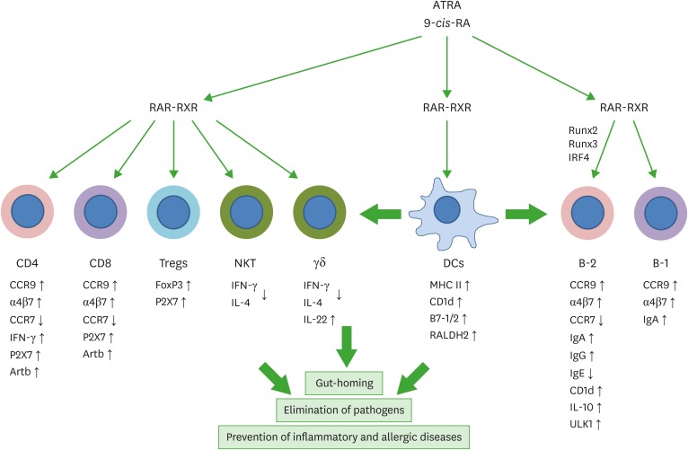
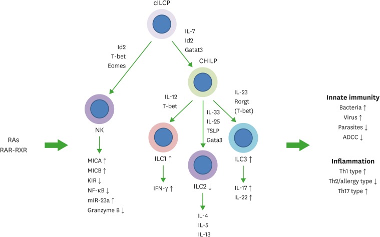
Lymphocytes, such as T cells, B cells, and innate lymphoid cells (ILCs), play central roles in regulating immune responses. Retinoic acids (RAs) are vitamin A metabolites, produced and metabolized by certain tissue cells and myeloid cells in a tissue-specific manner. It has been established that RAs induce gut-homing receptors on T cells, B cells, and ILCs. A mounting body of evidence indicates that RAs exert far-reaching effects on functional differentiation and fate of these lymphocytes. For example, RAs promote effector T cell maintenance, generation of induced gut-homing regulatory and effector T cell subsets, antibody production by B cells, and functional maturation of ILCs. Key functions of RAs in regulating major groups of innate and adaptive lymphocytes are highlighted in this article.

Keywords: B-cells; Innate lymphoid cells; NK cells; Retinoic acid; T-cells.


Conflict of interest statement

Conflict of Interest: The author declares no potential conflicts of interest.

Figures

Figure 1
Figure 1
Regulation of T cells and B cells by RAs. T cells and B cells express RARs and are major targets of RA regulation. RAs and their receptors appear to regulate T and B cells through genomic and non-genomic functions. RAs affect the effector function, gut-homing receptor expression, and apoptosis of CD4+ T cells. In the intestine, RAs promote gut-homing effector T cells (Th1 and Th17) and Tregs. Regulation of T cells by RAs occurs at the time of antigen priming, and DCs express RA-producing RALDH2. Moreover, RAs induce co-stimulatory receptors and RALDH2 in DCs. Therefore, DCs and other antigen presenting cells play central roles in regulating T cells by activating lymphocytes and producing RAs. RAs also induce the expression of P2X7 and Art2b on T cells to induce apoptosis caused by inflammatory mediators such as NAD and ATP, which are typically leaked out of dying cells in inflammatory conditions. This function of RAs induces effector T cell contraction in the intestines. RAs also induce gut-homing IgA-producing B-1 and B-2 cells in gut-associated lymphoid tissues. However, RAs function to suppress IgE production. RAs promote IL-10-producing regulatory T and B cells. The arrows indicate either positive (↑) or negative (↓) effect of RAs. RXR, retinoid X receptor; ULK1, UNC51-like kinase-1; IRF4, interferon regulatory factor 4.
Figure 2
Figure 2
Regulation of NK cells and ILCs by RAs. Common ILC progenitors (cILCP or also called αLP) and common helper-like ILC progenitors generate ILCs. Generation and functional maturation of innate lymphocytes are primarily regulated by cytokines and transcription factors. In addition, RAs regulate these NK cells and ILCs. RAs bidirectionally regulate NK cell functions. They induce MICA and MICB to indirectly activate NK cells but can directly suppress NK cell activation as well. While the overall effect of RAs on NK cells during various types of immune responses needs to be established, it appears that RAs function to support NK cell activity but restrain their activity in certain conditions. In general, RAs promote ILC1 and ILC3 activity, whereas they suppress ILC2 generation from ILC progenitors from the bone marrow or embryonic hematopoietic organs. RAs promote IL-22 production by ILC3 to strengthen the barrier immunity, and support IFN-γ production by ILC1. These ILC-boosting functions of RAs were observed on human ILCs as well. However, there appear to be species-specific differences in the regulation. Overall, RAs are important regulators of ILCs. The arrows indicate either positive (↑) or negative (↓) effect of RAs. ADCC, antibody-dependent cell cytotoxicity; KIR, killer inhibitory receptor; MICA, major histocompatibility complex class I chain-related protein A; MICB, major histocompatibility complex class I chain-related protein B; RXR, retinoid X receptor; TSLP, thymic stromal lymphopoietin.

References

    1. Fan X, Rudensky AY. Hallmarks of tissue-resident lymphocytes. Cell. 2016;164:1198–1211. - PMC - PubMed
    1. Masopust D, Schenkel JM. The integration of T cell migration, differentiation and function. Nat Rev Immunol. 2013;13:309–320. - PubMed
    1. Carbone FR. Tissue-resident memory T cells and fixed immune surveillance in nonlymphoid organs. J Immunol. 2015;195:17–22. - PubMed
    1. Hardy RR, Hayakawa K. Perspectives on fetal derived CD5+ B1 B cells. Eur J Immunol. 2015;45:2978–2984. - PMC - PubMed
    1. Clark MR, Mandal M, Ochiai K, Singh H. Orchestrating B cell lymphopoiesis through interplay of IL-7 receptor and pre-B cell receptor signalling. Nat Rev Immunol. 2014;14:69–80. - PMC - PubMed