

Ectopically Expressed Membrane-bound Form of IL-9 Exerts Immune-stimulatory Effect on CT26 Colon Carcinoma Cells
- PMID: 29503742
- PMCID: PMC5833119
- DOI: 10.4110/in.2018.18.e12
Ectopically Expressed Membrane-bound Form of IL-9 Exerts Immune-stimulatory Effect on CT26 Colon Carcinoma Cells
Abstract
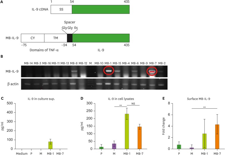
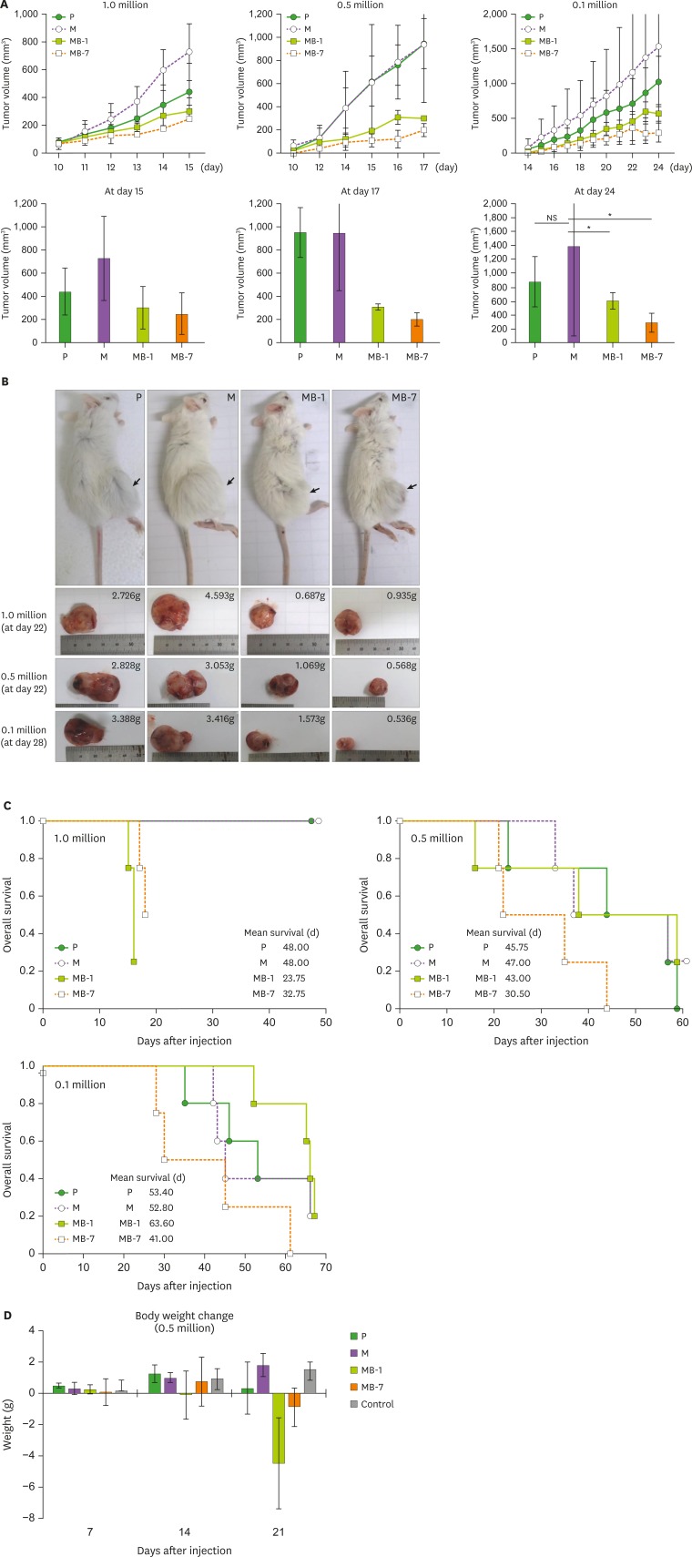
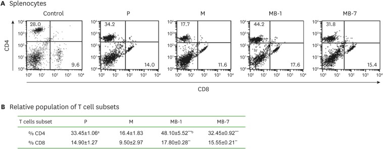
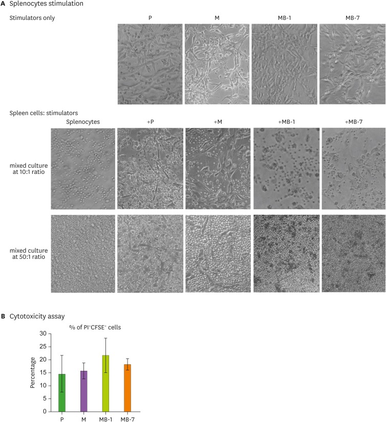
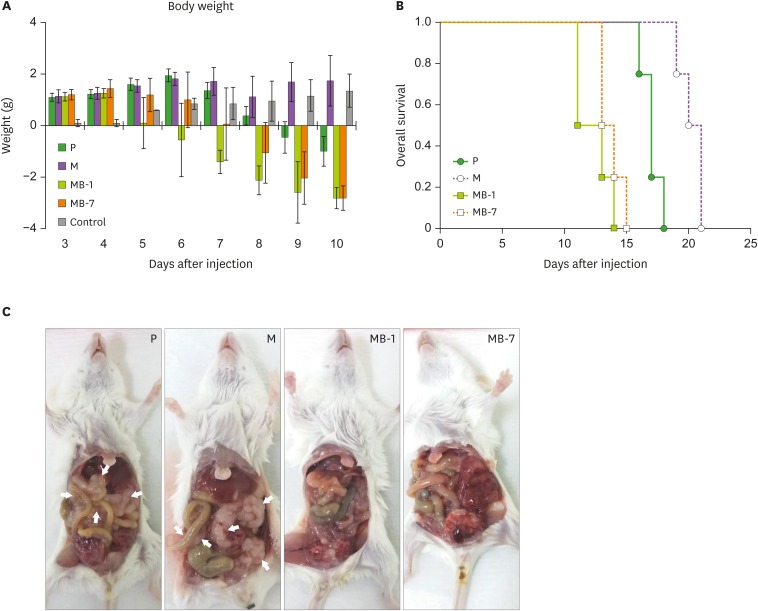
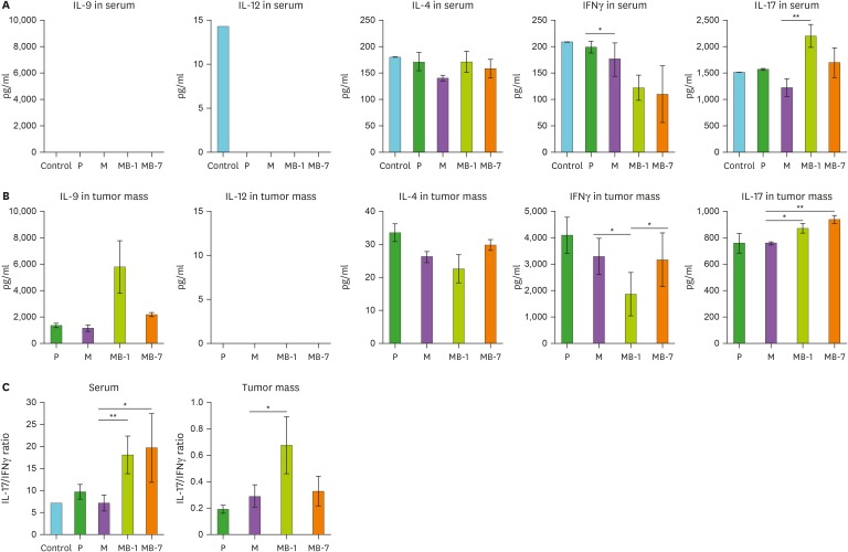
IL-9 is a known T cell growth factor with pleiotropic immunological functions, especially in parasite infection and colitis. However, its role in tumor growth is controversial. In this study, we generated tumor clones expressing the membrane-bound form of IL-9 (MB-IL-9) and investigated their influences on immune system. MB-IL-9 tumor clones showed reduced tumorigenicity but shortened survival accompanied with severe body weight loss in mice. MB-IL-9 expression on tumor cells had no effect on cell proliferation or major histocompatibility complex class I expression in vitro. MB-IL-9 tumor clones were effective in amplifying CD4+ and CD8+ T cells and increasing cytotoxic activity against CT26 cells in vivo. We also observed a prominent reduction in body weights and survival period of mice injected intraperitoneally with MB-IL-9 clones compared with control groups. Ratios of IL-17 to interferon (IFN)-γ in serum level and tumor mass were higher in mice implanted with MB-IL-9 tumor clones than those observed in mice implanted with control cells. These results indicate that the ectopic expression of the MB-IL-9 on tumor cells exerts an immune-stimulatory effect with toxicity. To exploit its benefits as a tumor vaccine, a strategy to control the toxicity of MB-IL-9 tumor clones should be developed.
Keywords: Cancer vaccine; Colon carcinoma; Interleukin-9; Membrane-bound; Toxicity.
Conflict of interest statement
Conflict of Interest: The authors declare no potential conflicts of interest.
Figures
Similar articles
-
Tumor cells ectopically expressing the membrane-bound form of IL-7 develop an antitumor immune response efficiently in a colon carcinoma model.Mol Cells. 2025 Feb;48(2):100175. doi: 10.1016/j.mocell.2024.100175. Epub 2024 Dec 30. Mol Cells. 2025. PMID: 39743142 Free PMC article.
-
The Membrane-Bound Form of IL-17A Promotes the Growth and Tumorigenicity of Colon Cancer Cells.Mol Cells. 2016 Jul;39(7):536-42. doi: 10.14348/molcells.2016.0048. Epub 2016 Jun 24. Mol Cells. 2016. PMID: 27378226 Free PMC article.
-
Membrane-bound p35 Subunit of IL-12 on Tumor Cells is Functionally Equivalent to Membrane-bound Heterodimeric Single Chain IL-12 for Induction of Anti-tumor Immunity.Immune Netw. 2016 Oct;16(5):305-310. doi: 10.4110/in.2016.16.5.305. Epub 2016 Oct 25. Immune Netw. 2016. PMID: 27799876 Free PMC article.
-
Heterogeneity in lymphokine profiles of CD4+ and CD8+ T cells and clones activated in vivo and in vitro.Immunol Rev. 1991 Oct;123:85-114. doi: 10.1111/j.1600-065x.1991.tb00607.x. Immunol Rev. 1991. PMID: 1684785 Review.
-
T-cell clones that react against autologous human tumors.Immunol Rev. 1990 Aug;116:33-62. doi: 10.1111/j.1600-065x.1990.tb00803.x. Immunol Rev. 1990. PMID: 2146210 Review.
Cited by
-
Th9 and Th17 Cells in Human Ulcerative Colitis-Associated Dysplastic Lesions.Clin Med Insights Oncol. 2024 Dec 7;18:11795549241301358. doi: 10.1177/11795549241301358. eCollection 2024. Clin Med Insights Oncol. 2024. PMID: 39651422 Free PMC article.
-
Interleukin-9 Inhibits Lung Metastasis of Melanoma through Stimulating Anti-Tumor M1 Macrophages.Mol Cells. 2020 May 31;43(5):479-490. doi: 10.14348/molcells.2020.0047. Mol Cells. 2020. PMID: 32326670 Free PMC article.
-
Tumor cells ectopically expressing the membrane-bound form of IL-7 develop an antitumor immune response efficiently in a colon carcinoma model.Mol Cells. 2025 Feb;48(2):100175. doi: 10.1016/j.mocell.2024.100175. Epub 2024 Dec 30. Mol Cells. 2025. PMID: 39743142 Free PMC article.
-
TH9, TH17, and TH22 Cell Subsets and Their Main Cytokine Products in the Pathogenesis of Colorectal Cancer.Front Oncol. 2019 Oct 4;9:1002. doi: 10.3389/fonc.2019.01002. eCollection 2019. Front Oncol. 2019. PMID: 31637216 Free PMC article. Review.
-
IL-9 and IL-9-producing cells in tumor immunity.Cell Commun Signal. 2020 Mar 30;18(1):50. doi: 10.1186/s12964-020-00538-5. Cell Commun Signal. 2020. PMID: 32228589 Free PMC article. Review.
References
-
- Renauld JC, van der Lugt N, Vink A, van Roon M, Godfraind C, Warnier G, Merz H, Feller A, Berns A, Van Snick J. Thymic lymphomas in interleukin 9 transgenic mice. Oncogene. 1994;9:1327–1332. - PubMed
-
- Veldhoen M, Uyttenhove C, van Snick J, Helmby H, Westendorf A, Buer J, Martin B, Wilhelm C, Stockinger B. Transforming growth factor-beta ‘reprograms’ the differentiation of T helper 2 cells and promotes an interleukin 9-producing subset. Nat Immunol. 2008;9:1341–1346. - PubMed
LinkOut - more resources
Full Text Sources
Other Literature Sources
Research Materials
Miscellaneous
