. 2018 Feb 27:6:e4383.
doi: 10.7717/peerj.4383. eCollection 2018.

Anther development in tribe Epidendreae: orchids with contrasting pollination syndromes

Affiliations

Anther development in tribe Epidendreae: orchids with contrasting pollination syndromes

Benjamín Valencia-Nieto et al. PeerJ. .

Abstract

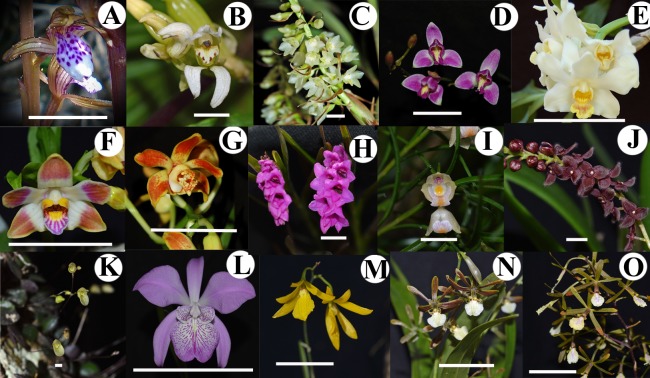
Background: Epidendreae is one of the most diverse tribes among the orchids with remarkable variation in life form, floral morphology and pollination syndromes. Its circumscription was recently revised and subtribes Agrostophyllinae and Calypsoinae were transferred into this tribe. One of the principal floral characters utilized in classification of orchids is the incumbency or bending of the column. This study records and compares late stages of anther, column and lip development, and discusses anther characters in fifteen representative taxa of five of the six subtribes in Epidendreae with respect to classification and pollination biology.

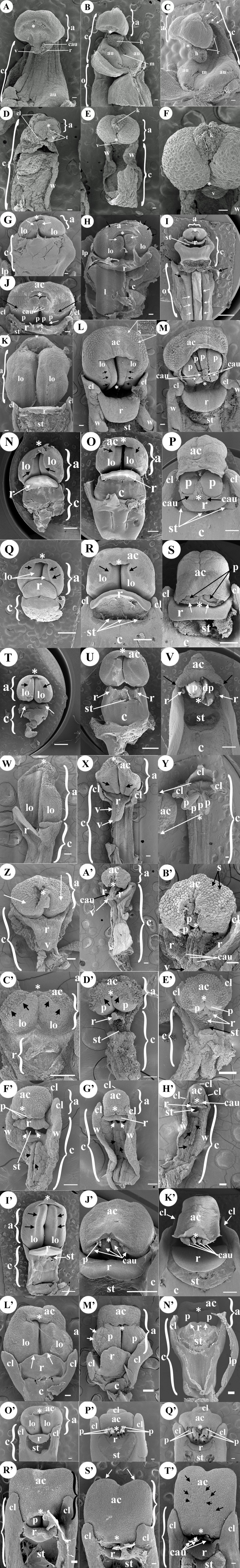
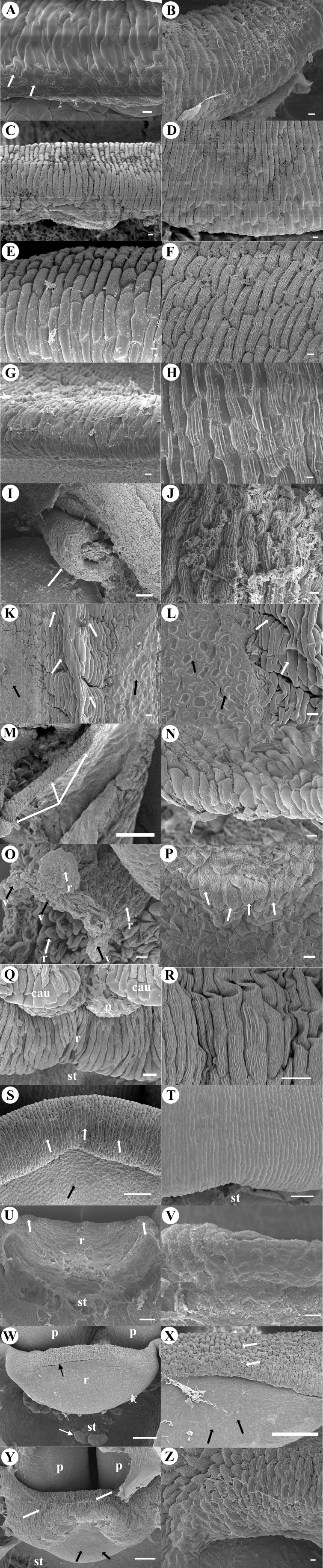
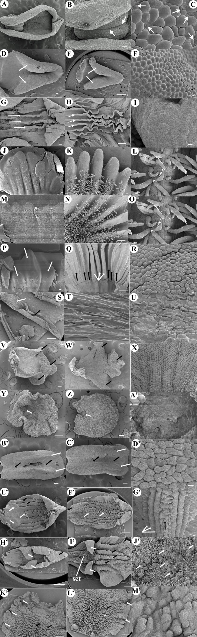
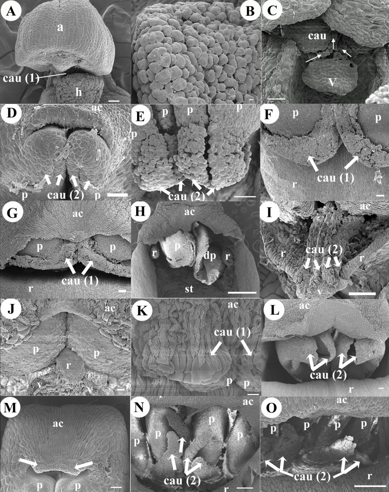
Methods: A series of late floral stages were sampled and fixed for examination under scanning electron microscope.

Results: Anther incumbency or bending in this group varies from 90° to almost 180°. Incumbency in the late stages of development is reached in Bletiinae, Ponerinae, Pleurothallidinae and Laeliinae whereas incumbency is reached early in its development in Corallorhiza and Govenia of Calypsoinae.

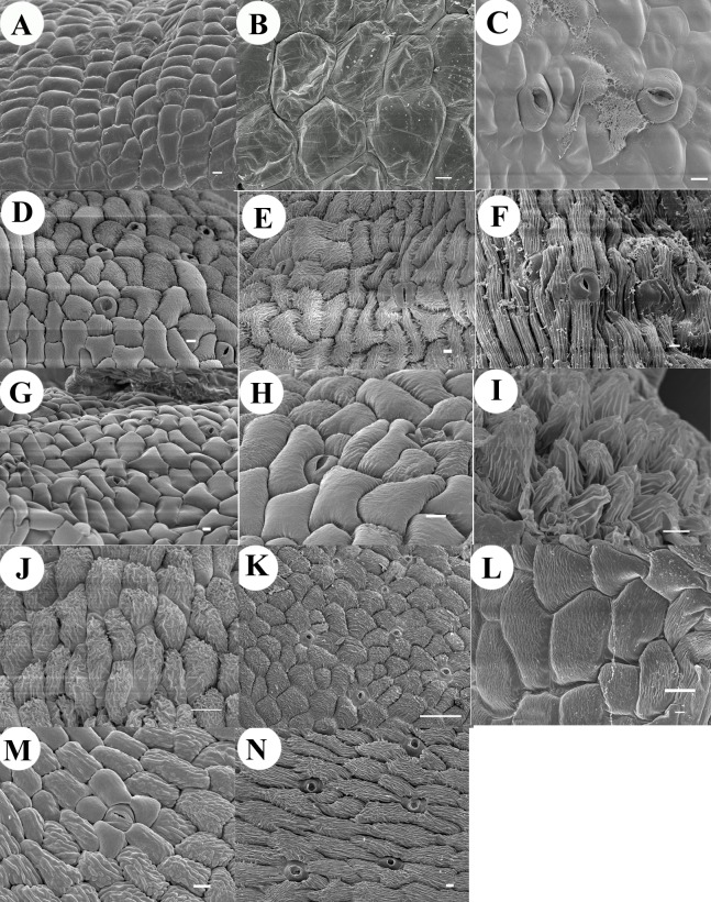
Discussion: Our observations indicate that the position of Chysis in subtribe Bletiinae needs revision based on differences in a number floral, and in particular of anther characters; and that Coelia only shares the early anther incumbency with Calypsoinae members, but not the rest of floral and anther characters. Anatomical characters such as crystals around the actinocytic stomata on the anther cap and sugar crystals in Laeliinae; lack of rostellum in Bletiinae; coalescent anther with the column, lack of trichomes and papillae on lip keels, and underdeveloped rostellum in Chysis; a mechanism by which the anther cap comes off (it is joined with the grooved lip by a claw) in Isochilus are all related to pollination syndromes and reproductive biology.

Keywords: Anther; Chysis; Development; Epidendreae; Incumbency; Orchidaceae; Pollination.


Conflict of interest statement

Victoria Sosa is an Academic Editor for PeerJ.

Figures

Figure 1
Figure 1. Studied species of Tribe Epidendreae.
(A) Corallorhiza maculata (Calypsoinae). (B) Govenia alba (Calypsoinae). (C) Coelia triptera (Coeliinae). (D) Bletia purpurea (Bletiinae). (E) Chysis bractescens (Bletiinae). (F) Chysis limminghei (Bletiinae). (G) Chysis laevis (Bletiinae). (H) Isochilus major (Ponerinae). (I) Ponera juncifolia (Ponerinae). (J) Stelis ciliaris (Pleurothallidinae). (K) Specklinia digita le (Pleurothallidinae). (L) Laelia speciosa (Laeliinae). (M) Oestlundia ligulata (Laeliinae). (N) Prosthechea squalida (Laeliinae). (O) Encyclia microbulbon (Laeliinae). Scale Bars: A = 1.5 cm. B = 1 cm. C = 1.2 cm. D = 5 cm. E = 9 cm. F = 5.5 cm. G = 6.5 cm. H = 1 cm. I = 5 mm. J = 4.5 mm. K = 3 mm. L = 14 cm. M = 1.4 cm. N = 2 cm. O = 3.5 cm.
Figure 2
Figure 2. Development micrographs of anther.
Late stages of development (column and anther) of Corallorhiza maculata (A, B, C). (A) Early incumbent anther. (B) Grown incumbent anther. (C) Lateral view bent over itself column and the incumbent anther. Govenia alba (D, E, F) (D) Early incumbent anther, column erect (same width throughout its length) and small developing wings. (E) Grown-up noticeably inflected anther. (F) Prominent keel in the middle of anther, just below the apical part of tegula (viscidium shield). Coelia triptera (G, H, I, J) (G) Early incumbent anther (H) Intermediate late stage incumbent anther. (I) Anther totally inflected, column hexagon-shaped, base joins the ovary which has pointed projections (black arrows) and with large grooves that cross longitudinally (white arrows). (J) Close-up of anther at anthesis, black arrows point the limit between anther cap and each pollinium respectively (two pairs of granulose caudicles exposed at apex). Bletia purpurea (K, L, M); (K) Early erect anther, slightly sub quadrate, bilobate with a trapezoid apex. (L) Intermediate late stage, anther inflected, the cucullate anther cap with actinocytic stomata embedded in its upper part (white arrows, close-up in the inset and Fig. 4B). (M) Anthesis, anther inflected, anther cap fully developed, pollinia (four upper visible), caudicles in the apex, at sides column wings. Chysis bractescens (N, O, P) (N) Early anther, erect, round shaped, bilobate with longitudinal lines marked on its epidermal tissue (black arrows). (O) Intermediate late stage anther, more quadrate and slightly less erect (apex with some degree of inflection), anther cap visible. (P) Galeate anther totally inflected, cucullate anther cap. A pair of pollinia exposed, with two laminar caudicles (one pair, black arrows) which surround the pollinia. Chysis limminghei (Q, R, S); (Q) Early anther, erect, round, bilobate with longitudinal lines marked on its epidermal tissue (black arrows). (R) Intermediate late stage anther, more quadrate, slightly less erect (apex with some degree of inflection), cucullate anther cap visible. (S) Galeate anther, totally inflected, cucullate anther cap. A pair of pollinia exposed with two laminar caudicles (one pair, white arrows). Chysis laevis (T, U, V); (T) Early erect anther, apex pronounced (trapezoid shaped), half round shaped, bilobate with longitudinal lines marked on its epidermal tissue (black arrows). Two very inconspicuous rostellum primordia (white arrows). (U) Intermediate late stage anther, slightly less erect (apex with some degree of inflection), bilobate, anther cap visible. Rostellum appendages vestiges underdeveloped (white arrows). (V) Incumbent anther, the anther cap conical shaped (like a hood), coalescent (fused by the sides to the column), dehiscent pollinia, open, without caudicles. No rostellum developed, only two appendixes underdeveloped and rolled on themselves (white arrows). Isochilus major (W, X, Y); (W) Early erect anther, semi-rectangular vertical with a pointed apex, biloculate. Column narrow and slender. (X) Intermediate late stage anther, semi rectangular shaped, pointed apex of conspicuous thickened cells, biloculate, each locule with longitudinal division (black arrow). (Y) Four elliptical inflected pollinia. Anther cap was not observed in this stage because it was lost during dissection; the lone structure (anther cap) was placed and observed at the side of the column in the slide (white arrows). Ponera juncifolia (Z, A′, B′); (Z) Early erect anther, rounded and bilobed, each lobule ovoid (white arrows). (A′) Intermediate late stage anther with caudicles exposed. (B′) Anther cap very conspicuous with actinocytic stomata embedded (black arrows). Two pollinia visible in each locule, four long caudicles exposed. Stelis ciliaris (C′, D′, E′) (C′) Early anther erect bilobed, each lobule ovoid with a small rounded apex, anther cap beginning its differentiation from the tissue of the rest of the anther, a clearly marked line delimits it (black arrows). (D′) Intermediate late stage anther, bilobed, anther cap clearly marked (black arrows) below two ovoid pollinia in close contact to upper part of rostellum tip (bifid viscidium, white arrow). (E′) Incumbent anther, anther cap with inverted heart shape, two pollinia exposed, in close contact to the tip of rostellum the bifid viscidium with abundant secretions. Specklinia digitale (F′, G′, H′) (F′) Inflected anther, cucullate, tip of two pollinia exposed. Column beneath with two teeth (white arrows). Below column narrows and canaliculated (black arrows). Column wings differentiated. (G′) Anther slightly more inflected, column with two teeth (white arrows). (H′) Inflected cucullate anther, two caudicles exposed, column is slim and long, canaliculated in the middle portion (black arrows), and with vertical wings. Laelia speciosa (I′, J′, K′) (I′) Erect anther, quadrate, bilobed, with a trapezoidal apex. Lobules with median longitudinal division each (black arrows). Column short and narrow. (J′) Intermediate late stage inflected anther, anther cap sub-globose with round striated cells. Underneath four caudicles visible. (K′) Inflected anther, the anther cap is oblong, with its apex folded up. Four large granulose caudicles are exposed in two pairs. Oestlundia ligulata (L′, M′, N′) (L′) Early erect anther, ovoid, bilobed, upper part by the apex a pleat of tissue is formed, early differentiation of the mid-part of the anther cap. (M′) Intermediate late stage inflected anther, bilobed with anther cucullate. (N′) Inflected anther, sub-cubic almost straight, two pollinia visible. Prosthechea squalida (O′, P′, Q′); (O′) Early erect anther, oblong and bilobed. (P′) Intermediate late stage inflected anther inflected the anther cap ellipsoid. Under anther cap four pollinia, two pairs of caudicles exposed (white arrows). (Q′) Inflected anther, anther cap truncate obovate shaped. Under it four pollinia, with four large, granulose caudicles in two pairs. Encyclia microbulbon (R′, S′, T′). (R′) Inflected anther quadrate with upper corners rounded, under anther cap two pollinia can be seen, (S′) More inflected anther, sub-quadrate with upper part bi-lobed and corners rounded (white arrows). At the apex of anther cap tip of two pollinia exposed. (T′) Inflected anther, obcordate semi-quadrate. Anther cap with abundant semi-actinocytic stomata (black arrows). Below anther cap, four pollinia are visible and two pairs of caudicles exposed. Scale Bars: A, B, C, D, E, F, G, H, J, K, L, M, W, X, Y, Z, A′, B′, C′, D′, E′, F′, G′, H′, L′, M′, N′, O′, P′, Q′, R′, S′ = 100 µm. I, N, O, P, Q, R, S, T, U, V, I′, J′, K′ = 1 mm. Abbreviations: *, anther apex; a, anther; ac, anther cap; ap, appendage; au, auricle; c, column; cau, caudicle; cl, clinandrium; dp, dehiscent pollinia; h, hamulus; l, lip; lo, lobule; lp, lateral petal; m, mentum; o, ovary; p, pollinium; r, rostellum; s, stomata; st, stigma; t, tegula; v, viscidium; w, column wings. Photo Credit SEM Images: Silvia Espinosa Matías. Figure edition: Benjamín Valencia-Nieto.
Figure 3
Figure 3. Rostellum micrographs.
Rostellum development of Coelia triptera (A, B). (A) Early stages, smooth rectangular cells, numerous intercellular connections (white arrows). (B) Rostellum at anthesis, wide barrier of rectangular cells. Bletia purpurea (C, D). (C) Early incipient rostellum. (D) Rostellum at anthesis (half-moon shape structure) of rectangular epidermal cells. Chysis limminghei (E, F). (E) Tip of early-rostellum with rectangular epidermal cells with striate cuticle. (F) Rostellum shield-like made up with rectangular epidermal cells arranged in palisade. Chysis bractescens (G, H). (G) Tip of projecting early-rostellum with rectangular epidermal cells. (H) Rostellum shield-like (rectangular epidermal cells in palisade). Chysis laevis (I, J). (I) Close-up of one of two appendixes of underdeveloped rostellum rolled on itself (white arrow). (J) Underdeveloped rostellum made up with striated rectangular epidermal cells arranged in palisade. Isochilus major (K, L). (K) Triangular rostellum with two cellular types: elongated cells (white arrows), and the glandular cellular type (black arrows) (L) Close-up of two cellular types of rostellum, the viscidium (black arrows). Ponera juncifolia (M, N). (M) Right side of v-shaped shield rostellum (white arrows) (N) Closer view of the border of rostellum with fibrous cells. Stelis ciliaris (O, P). (O) Tip of upper part of rostellum, the viscidium is bifid with very fibrous cells, in the middle the tissue is covered with a dense secretions (black arrows) (P) Rostellum with big round fibrous cells (white arrows). Specklinia digitale (Q, R). (Q) Rostellum with large longitudinal striated cells. (R) Close-up of rostellum. Laelia speciosa (S, T), (S) Tip of early rostellum with semi-rectangular cells (white arrows), the abaxial side with smooth irregular cells (black arrow). (T) Close-up of rostellum at anthesis. Oestlundia ligulata (U, V); (U) Transverse half-moon shaped rostellum. (V) Rostellum tip. Prosthechea squalida (W, X). (W) Early ellipsoid rostellum, border with thickened cells (black arrow two small bulges of stigma (white arrow). (X) Close-up of the superior part or rostellum (white arrows), lower part with smooth cells (black arrows). Encyclia microbulbon (Y, Z) (Y) Early pointy tongue-shaped rostellum with longitudinal elongated striated cells (white arrows), the abaxial side is smooth (black arrows). (V) Close-up of the striated cells at the tip of rostellum. Scale Bars: A, B, C, D, E, F, G, H, J, K, L, N, O, P, Q, R, S, T, Z = 10 µm. I, M, U, V, W, X, Y = 100 µm. Abbreviations: cau, caudicle; p, pollinium; r, rostellum; st, stigma; v, viscidium. Photo Credit SEM Images: Silvia Espinosa Matías. Figure edition: Benjamín Valencia-Nieto.
Figure 4
Figure 4. Micrographs of anther cap cuticle.
Cuticle of the epidermis of the anther cap of (A) Corallorhiza maculata with smooth cells without stomata. (B) Govenia alba with smooth to slightly wrinkled cells without stomata. (C) Coelia triptera with smooth cells showing actinocytic stomata and secretions. (D) Bletia purpurea with slightly striated cells, note the actinocytic stomata. (E) Chysis limminghei with conspicuous striated cells and actinocytic stomata. (F) Chysis bractescens with longitudinal striated cells and actinocytic stomata. (G) Isochilus major with smooth cells and actinocytic stomata. (H) Ponera juncifolia with smooth to slightly striated cells and actinocytic stomata. (I) Stelis ciliaris with striated slightly corrugated cells without stomata. (J) Specklinia digitale with densely striated cells without stomata. (K) Laelia speciose with round striated cells and actinocytic stomata. (L) Oestlundia ligulata with round slightly striated cells without stomata. (M) Prosthechea squalida with slightly wide striated cells and actinocytic stomata. (N) Encyclia microbulbon with conspicuous striated cells and semi-actinocytic stomata. Scale Bars: A–J, L–N = 10 µm, K = 100 µm. Photo Credit SEM Images: Silvia Espinosa Matías. Figure edition: Benjamín Valencia-Nieto.
Figure 5
Figure 5. Early and anthesis stages of lip development.
Corallorhiza maculata (A, B, C). (A) Lip. (B) Protuberances of lip forming a channel in the middle. (C) Close-up of the cells of the protuberances, presence of abundant pores. Coelia triptera (D, E, F). (D) Early lip development with two bulges (white arrows). (E) Lip at anthesis arrowhead shaped bulging sides (white arrows). (F) Smooth rounded single epidermis cells. Bletia purpurea (G, H, I). (G) Early stage lip (three central keels white arrows). (H) Anthesis lip (three central keels with white arrows and 3–4 lateral accessory keels developed with black arrows). (I) Epidermis cells. Chysis limminghei (J, K, L). (J) Early lip development, five central keels with large papillae epidermal cells protruding. (K) Lip at anthesis, five keels with abundant simple trichomes. (L) Close-up of trichomes. Chysis bractescens (M, N, O). (M) Early lip development, five central keels with large papillae epidermal cells protruding. (N) Lip at anthesis, five keels with abundant large simple trichomes. (O) Close-up of trichomes. Chysis laevis (P, Q, R). (P) Early lip development with three central keels prominent (white arrows). (Q) Base of the lip at anthesis, three central keels (white arrows), five accessory keels at each side of the lip (black arrows). (R) Close-up of epidermal cells of the keels. Isochilus major (S, T, U). (S) Lip at anthesis, flat channeled (white arrow), forming a slender channel (black arrow). (T) Close-up of epidermal cells of the flat part of lip. (U) Close-up of epidermis cells of the conduplicate part and lip apex. Ponera juncifolia (V, W, X). (V) Early lip development, tri-lobed (black arrows). (W) Lip at anthesis, mid-lobe apex slightly wrapped (black arrows). (X) Epidermis cells. Stelis ciliaris (Y, Z, A′). (Y) Early lip, at base hollow in the middle (white arrow). (Z) Lip at anthesis, hollow at base (white arrow) with trichomes. (A′) Close-up of hollow with trichomes. Specklinia digitale (B′, C′, D′). (B′) Early lip, two simple thickened keels (white arrow), a channel formed between them (black arrows). (C′) Lip at anthesis with two simple thickened keels (white arrows) and channel between (black arrows). (D′) Cells on the keels. Oestlundia ligulata (E′, F′, G′). (E′) Early lip with three central keels (white arrows), two accessory keels at each side (black arrows). (F′) Lip at anthesis unguiculate to the base (black arrow), central keels highly verrucose (white arrows). (G′) Central highly verrucose keel (white arrow) besides accessory keels black arrows). Prosthechea squalida (H′, I′, J′). (H′) Tri-lobed lip, the tip wrapped on itself (black arrow), two small lateral lobes are incurved (white arrows). (I′) Unguiculate lip at the base, tri-lobed, the lateral lobes incurved (white arrows), the central portion is composed of abundant glandular trichomes. From the center to the tip of the mid-lobe, three large central verrucose keels (black arrows) and two or more lateral keels are present. (J′) Central portion with abundant glandular trichomes secreting large amounts of sugar crystals (white arrows). Encyclia microbulbon (K′. L′. M′). (K′–L′) Mid-lobe lip, circular-ovate with crenulated margins (white arrows), the center the surface is very verrucose (black arrows) without keels. (M′) Close up of verrucose surface cells. Scale Bars: C, F, I, R, T, U, A′ = 10 µm, A, B, D, G, J, K, L, M, O, P, V, W, X, Y, Z, B′, C′, D′, E′, G′, J′, M′ =100 µm. E, H, N, Q, S, F′, H′, I′, K′, L′ = 1 mm. Photo Credit SEM Images: Silvia Espinosa Matías. Figure edition: Benjamín Valencia-Nieto.
Figure 6
Figure 6. Micrographs of pollinia and caudicles.
Pollinia and caudicles of (A) Corallorhiza maculata one caudicle connected to hamulus. (B) Corallorhiza maculata smooth, very rounded, granulose cells of the hamulus. (C) Govenia alba tegula composed by a strand of granulose cells with viscidium resembling a little shield, connected to the apex of the pollinia (one pair). (D) Coelia triptera, two pollinia and two pairs of caudicles with polyhedral cells. (E) Bletia purpurea, four upper pollinia and two pairs of caudicles with polyhedral cells. (F) Chysis bractescens, two pollinia and one pair of linear caudicles with membranaceous cells. (G) Chysis limminghei, two pollinia and one pair of linear caudicles with membranaceous cells. (H) Chysis laevis, two pollinia, one pollinium without caudicles and another dehiscent pollinium. (I) Ponera juncifolia, note two pairs of caudicles, pollinia covered with anther cap. (J) Stelis ciliaris, two pollinia and central bifid tip of rostellum. (K) Specklinia digitale, two pollinia and one pair of rectangular smooth caudicles. (L) Laelia speciosa with four large granulose caudicles exposed in two pairs. (M) Oestlundia ligulata, two pollinia and pleat of tissue in the apex (white arrows) of the anther cap. (N) Prosthechea squalida, four pollinia and two pairs of caudicles. (O) Encyclia microbulbon, four pollinia and two pairs of caudicles. Scale Bars: A, C, D, E, F, G, I, M, N, L = 100 µm. B, J, K = 10 µm. H =1 mm. Abbreviations: ac, anther cap; cau, caudicle (irregular mass of cells); cau (1), one pair of caudicles; cau (2), two pair of caudicles; h, hamulus; p, pollinium; r, rostellum; st, stigma; v, viscidium. Photo Credit SEM Images: Silvia Espinosa Matías. Figure edition: Benjamín Valencia-Nieto.

Similar articles

References

    1. Arditti J. Fundamentals of orchid biology. John Wiley & Sons; New York: 1992.
    1. Blanco MA, Barboza G. Pseudocopulatory pollination in Lepanthes (Orchidaceae: Pleurothallidinae) by fungus gnats. Annals of Botany. 2005;95:763–772. doi: 10.1093/aob/mci090. - DOI - PMC - PubMed
    1. Borba EL, Braga PIS. Biologia reprodutiva de Pseudolaelia corcovadensis (Orchidaceae): melitofilia e autocompatibilidade em uma Laeliinae basal. Revista Brasileira de Botânica. 2003;26:541–549. doi: 10.1590/S0100-84042003000400013. - DOI
    1. Chase MW, Cameron KM, Barrett RL, Freudenstein JV. DNA data and Orchidaceae systematics: a new phylogenetic classification. In: Dixon KW, Kell SP, Barret RL, Cribb PJ, editors. Orchid conservation. Natural History Publications; Kota Kinabalu, Sabah: 2003.
    1. Chase MW, Cameron KM, Freudenstein JV, Pridgeon AM, Salazar GA, Van den Berg C, Schuiteman A. An updated classification of Orchidaceae. Botanical Journal of the Linnean Society. 2015;177:151–174. doi: 10.1111/boj.12234. - DOI

LinkOut - more resources