Review
. 2018 Feb 6:6:2.
doi: 10.3389/fcell.2018.00002. eCollection 2018.

A Salutary Role of Reactive Oxygen Species in Intercellular Tunnel-Mediated Communication

Affiliations
Review

A Salutary Role of Reactive Oxygen Species in Intercellular Tunnel-Mediated Communication

Dacheng Liang. Front Cell Dev Biol. .

Abstract

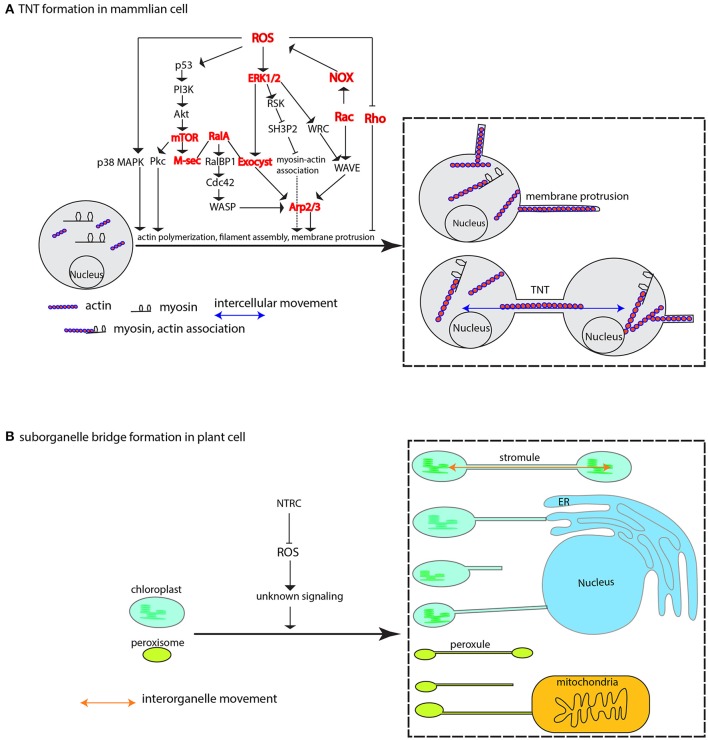
The reactive oxygen species, generally labeled toxic due to high reactivity without target specificity, are gradually uncovered as signaling molecules involved in a myriad of biological processes. But one important feature of ROS roles in macromolecule movement has not caught attention until recent studies with technique advance and design elegance have shed lights on ROS signaling for intercellular and interorganelle communication. This review begins with the discussions of genetic and chemical studies on the regulation of symplastic dye movement through intercellular tunnels in plants (plasmodesmata), and focuses on the ROS regulatory mechanisms concerning macromolecule movement including small RNA-mediated gene silencing movement and protein shuttling between cells. Given the premise that intercellular tunnels (bridges) in mammalian cells are the key physical structures to sustain intercellular communication, movement of macromolecules and signals is efficiently facilitated by ROS-induced membrane protrusions formation, which is analogously applied to the interorganelle communication in plant cells. Although ROS regulatory differences between plant and mammalian cells exist, the basis for ROS-triggered conduit formation underlies a unifying conservative theme in multicellular organisms. These mechanisms may represent the evolutionary advances that have enabled multicellularity to gain the ability to generate and utilize ROS to govern material exchanges between individual cells in oxygenated environment.

Keywords: ROS; intercellular movement; interorganelle transport; macromolecule movement; membrane protrusions; multicellularization; plasmodesmata; tunneling nanotubes (TNTs).


Figures

Figure 1
Figure 1
Symplastic dye movement is associated with ROS. The symplastic dye 10 kd F-Dextran moves freely at the heart stage of WT and ise2 mutant, but its movement is restricted at the torpedo stage due to the formation of symplastic domain. Higher ROS content in the ise2 mutant lifts this restriction. Green depicts F-Dextran (a). In the epidermal/trichome (e/t) boundary, LYHC dye moves freely between the trichome cells, but cannot cross the e/t boundary into epidermal cell. Sodium azide, a known ROS-inducing agent can break this blockage. Yellow color depicts the LYCH. (c–e) A root-to-shoot silencing system (RtSS) demonstrates the long-distance movement of small RNA-mediated gene silencing movement. (c) An RtSS plant shows the silencing pattern after 15-day Dex induction. (d,e) Longitudinal sectioning of RtSS has shown that silencing front moves from the root to the shoot (d), but the mutation in type III peroxidase RCI3 (R145K) heavily retards the movement (e). (a,b) were drawn according to the description by Kim I. et al. (2002) and Christensen et al. (2009) respectively. (e) was reprinted from Liang et al. (2014) under a Creative Commons Attribution License granted to Peer J.
Figure 2
Figure 2
Peroxidase-catalyzed cell wall remodeling dictates PD transport. In the apoplastic space (light yellow), type III peroxidase (POX) performs peroxidative cycle (blue arrows) and oxidative cycle (green arrows) to regulate H2O2 and superoxide/O2 level. Apoplastic ROS from the two cycles lead to cell wall tightening or loosening by crosslinking or depolymerizing cell wall components, thereby shrinking or enlarging the plasmodesmatal passage. Apoplastic H2O2 can also, through aquaporins-mediated transmembrane transport, enter the cytoplasm to initiate downstream signaling events, e.g., MAPK cascade signaling and cytoskeletal remodeling, resulting in membranous protrusion (see the section ROS is required to form intercellular/interorganelle bridge for the details). ROS imbalance inflicted by mutation in ISE1, ISE2, GAT1 localized in the subcellular compartments also impacts on intercellular transport. It's currently unknown whether or how ROS from subcellular compartments contribute to apoplastic ROS pool. Protein that moves through PD sleeve may be subject to ER stress-induced ROS, thus requires a post-movement refolding (details in the text). The peroxidase/oxidase cycling was adapted from Berglund et al. (2002) with the permission from Rightslink®.
Figure 3
Figure 3
Protein unloading from phloem to surrounding cells in root is associated with redox homeostasis. GFP is specifically expressed in the companion cell under the control of AtSuc2 promoter. It's not retained in the companion cell but rather disperses into sieve element (SE) and other parts of roots including stele, cortex, RAM (round circle), and root cap (A). mRNA of GFP is less likely to move into surrounding cells to be translated into fluorescence signal as plants harboring either bigger size of GFP or subcellular targeted GFP only show green fluorescence in the companion cells. Loss-of-function mutation in thioredoxin gene GAT1 for redox regulation (B), or gain-of-function mutation in callose synthase gene Cals3 shows defective GFP trafficking (C). (A–C) were drawn according to the description by Imlau et al. (1999), Benitez-Alfonso et al. (2009), Vatén et al. (2011) respectively.
Figure 4
Figure 4
Intercellular KN1 movement through PD requires a post-movement refolding, which could result from ER stress. The non-cell autonomous feature of KN1C (trafficking domain of KN1) protein renders the GFP-GL1-KN1C fusion as a movement protein that moves from mesophyll cell to epidermal cell, whence the GL1 part in the fusion initiates the developmental program for trichome specification. Chaperonin CCT8 is required in the destination cell, in this case the epidermal cell, to refold the post-movement protein. This figure was drawn based on the description by Xu et al. (2011).
Figure 5
Figure 5
ROS-induced formation of membrane protrusion, intercellular and interorganelle bridge. (A) At physiologically higher ROS levels, TNTs are formed between cells via cytoskeleton-based (e.g., actin, myosin) membrane protrusion. ROS-induced diversified pathways including p38 MAPK, PI3k-Akt-mTOR signaling, ERK1/2 signaling, Rho GTPases family, and M-Sec-RalA-Exocyst complex, are shown to promote membrane protrusions mainly via Arp2/3-mediated actin cytoskeletal remodeling. The cooperative association between myosin and actin that is activated by ERK signaling plays an important role in mobilizing various related components and mediating membrane-cytoskeleton coordination. (B) In plant cells, the membrane-encircled organelles, e.g., the chloroplast and peroxisome, can form inter-organellar bridge between the same type of organelle (the stromule and peroxule) and different types of organelles under higher ROS condition. Repression of NTRC leads to increased stromules. The signaling mechanism is currently unknown. Arrows may not denote direct activation. Several key nodes in the signaling networks are colored in red, and Arp1/2 complex, Exocyst complex, NOX, ERK, p38 MAPK, Rac and Rho are also encoded in plant genomes. p38 MAPK, p38 mitogen-activated protein kinase; Pkc, protein Kinase C; PI3k, phosphatidylinositol-3-kinase; AKT, protein kinase B; mTOR, mammalian target of rapamycin; M-Sec, Myeloid and M cells-expressing Sec6 homolog, also known as TNF alpha-induced protein 2, or Primary response gene B94; RalA, Ras-related protein Ral-A; RalBP1, RalA-binding protein 1; Cdc42, Cell division control protein 42 homolog; WASP, the Wiskott–Aldrich Syndrome protein; ERK1/2, extracellular signal-regulated kinase 1; Exocyst, an octameric complex; RSK, ribosomal S6 kinase; SH3P2, Src homology-3 (SH3) domain-containing protein 2; Arp2/3, actin-related complex 2/3; WRC, WAVE2 Regulatory Complex; NOX, NADPH oxidase; WAVE, WASP family verprolin-homologous protein; NTRC, NADP-thioredoxin reductase C.
Figure 6
Figure 6
A tentative model to depict the relationship among cell wall remodeling, membrane protrusion, complex PD formation and increased ROS level in plant cells. Plasmodesmata (PD)-mediated intercellular transport is usually regulated by PD-localized proteins (PDLPs) and other PD-associated regulators (e.g., callose synthase, glucanase), and involves no cell wall remodeling (A). In apoplast, ROS-generating enzymes including type III peroxidase (POX) and other cell wall-localized oxidases drive cell wall remodeling. Apoplastic H2O2 increment and subsequent conversion into hydroxyl radical from H2O2 leads to cell wall loosening via oxidative scission of cell wall polysaccharides and ROS-activating enzymatic breakdown of cell wall-associated proteins. PD embedded in the loosened cell wall expands (B), resulting in increased intercellular exchange. In the symplast, H2O2-induced protruding membrane may cross the loosened cell wall to form novel PD or fuse with existing PD to form complex PD (C). However, too high ROS cause either cell death (apoptosis) or middle lamella dissolution, resulting in the physical cut-off between cells (D).

Similar articles

Cited by

References

    1. Abid M. R., Kachra Z., Spokes K. C., Aird W. C. (2000). NADPH oxidase activity is required for endothelial cell proliferation and migration. FEBS Lett. 486, 252–256. 10.1016/S0014-5793(00)02305-X - DOI - PubMed
    1. Abounit S., Bousset L., Loria F., Zhu S., de Chaumont F., Pieri L., et al. . (2016). Tunneling nanotubes spread fibrillar α-synuclein by intercellular trafficking of lysosomes. EMBO J. 35, 2120–2138. 10.15252/embj.201593411 - DOI - PMC - PubMed
    1. Adamec L. (2006). Respiration and photosynthesis of bladders and leaves of aquatic utricularia species. Plant Biol. 8, 765–769. 10.1055/s-2006-924540 - DOI - PubMed
    1. Adamec L. (2007). Oxygen concentrations inside the traps of the carnivorous plants Utricularia and Genlisea (Lentibulariaceae). Ann. Bot. 100, 849–856. 10.1093/aob/mcm182 - DOI - PMC - PubMed
    1. Agustí J., Gimeno J., Merelo P., Serrano R., Cercós M., Conesa A., et al. . (2012). Early gene expression events in the laminar abscission zone of abscission-promoted citrus leaves after a cycle of water stress/rehydration: involvement of CitbHLH1. J. Exp. Bot. 63, 6079–6091. 10.1093/jxb/ers270 - DOI - PMC - PubMed

LinkOut - more resources