Review
. 2018 Jan 9:2018:5610347.
doi: 10.1155/2018/5610347. eCollection 2018.

Fatty Images of the Heart: Spectrum of Normal and Pathological Findings by Computed Tomography and Cardiac Magnetic Resonance Imaging

Affiliations
Review

Fatty Images of the Heart: Spectrum of Normal and Pathological Findings by Computed Tomography and Cardiac Magnetic Resonance Imaging

Giuseppe Cannavale et al. Biomed Res Int. .

Abstract

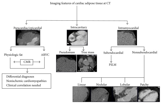
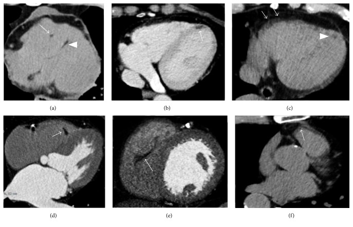
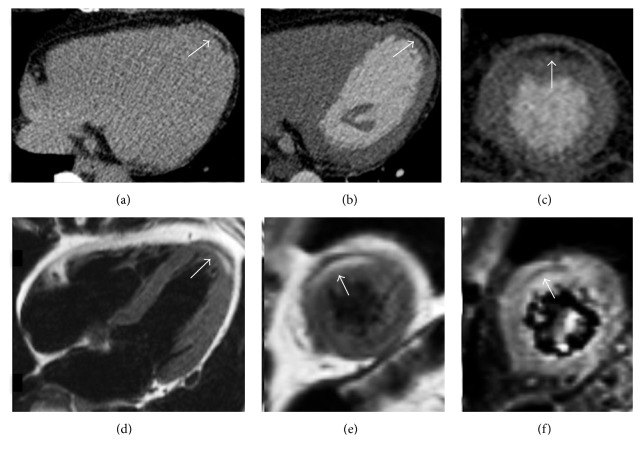
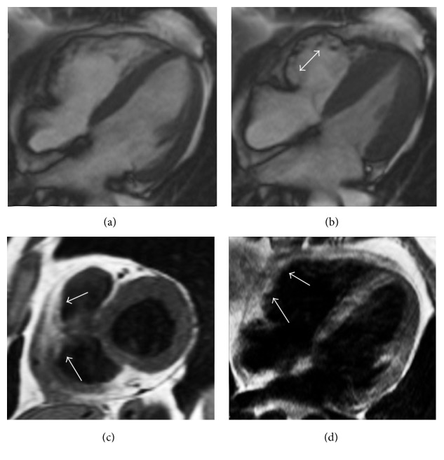
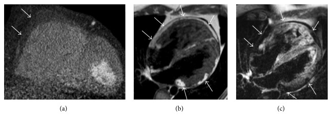
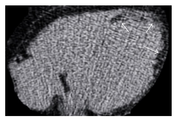
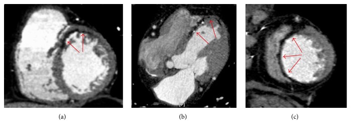
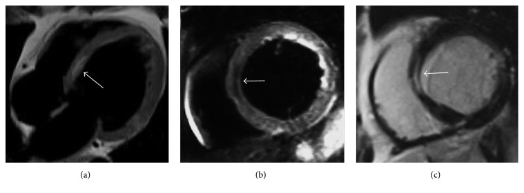
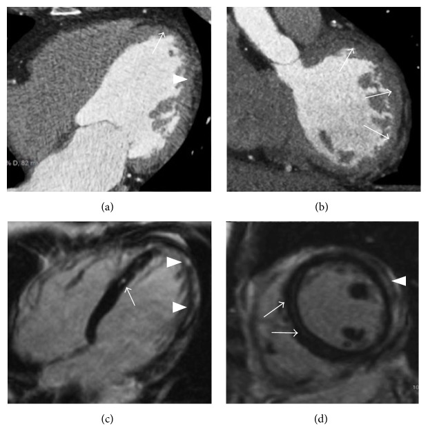
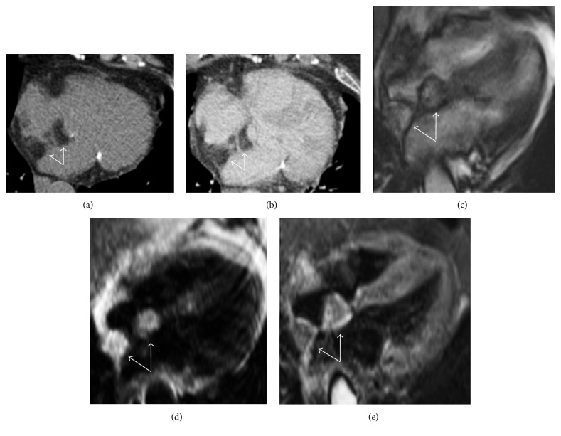
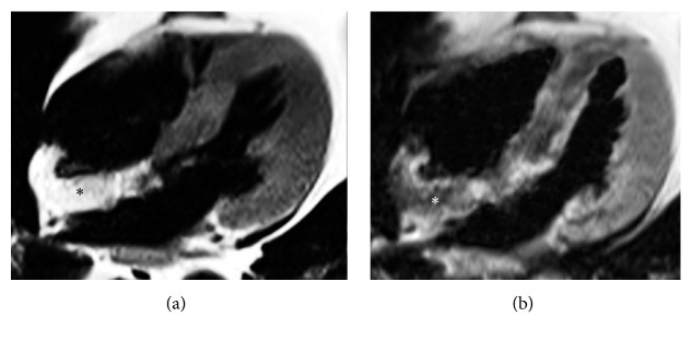
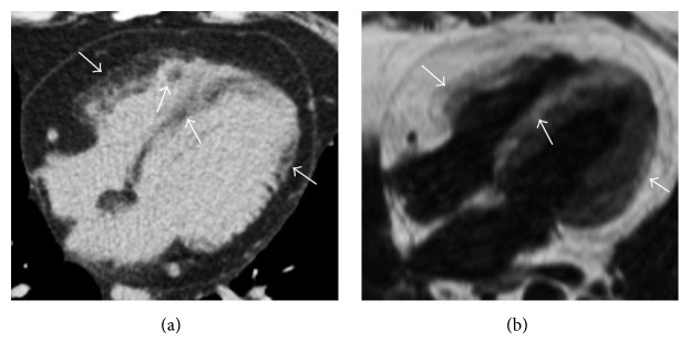
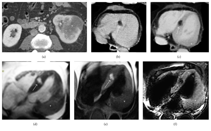
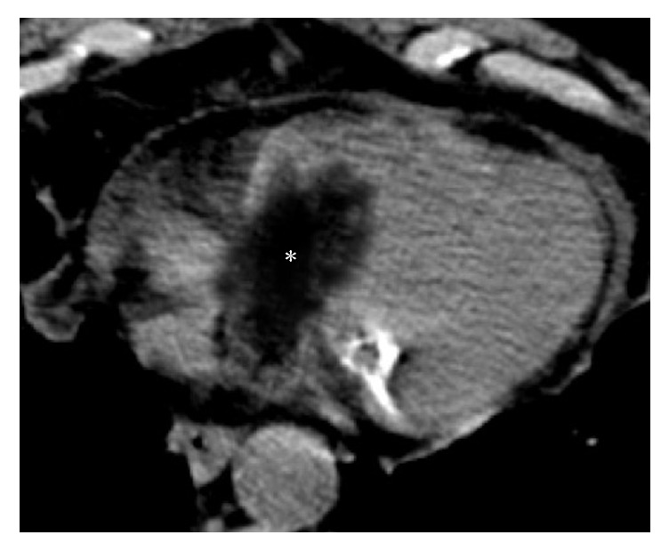
Ectopic cardiac fatty images are not rarely detected incidentally by computed tomography and cardiac magnetic resonance, or by exams focused on the heart as in general thoracic imaging evaluations. A correct interpretation of these findings is essential in order to recognize their normal or pathological meaning, focusing on the eventually associated clinical implications. The development of techniques such as computed tomography and cardiac magnetic resonance allowed a detailed detection and evaluation of adipose tissue within the heart. This pictorial review illustrates the most common characteristics of cardiac fatty images by computed tomography and cardiac magnetic resonance, in a spectrum of normal and pathological conditions ranging from physiological adipose images to diseases presenting with cardiac fatty foci. Physiologic intramyocardial adipose tissue may normally be present in healthy adults, being not related to cardiac affections and without any clinical consequence. However cardiac fatty images may also be the expression of various diseases, comprehending arrhythmogenic right ventricular dysplasia, postmyocardial infarction lipomatous metaplasia, dilated cardiomyopathy, and lipomatous hypertrophy of the interatrial septum. Fatty neoplasms of the heart as lipoma and liposarcoma are also described.


Figures

Figure 1
Figure 1
Imaging features of cardiac adipose tissue by computed tomography. CT: computed tomography; ARVC: arrhythmogenic right ventricular dysplasia; CMR: cardiac magnetic resonance; PILM: postmyocardial infarction lipomatous metaplasia.
Figure 2
Figure 2
Typical incidental physiologic cardiac fatty images detected by CT. (a) Small fatty foci are seen in right ventricular moderator band (arrow) as within the interventricular septum (arrowhead) and in the left ventricular myocardial apex ((b) arrow). A small amount of pericardial fluid is also noted in (a). (c) A linear shaped adipose infiltration is, respectively, illustrated in the right ventricular free wall (arrows) as in the left ventricular apex (arrowhead), within the right ventricular moderator band ((d) arrow), in the right ventricular trabeculae ((e) arrow), and at the right ventricular outflow tract ((f) arrow).
Figure 3
Figure 3
Thoracic noncontrast CT axial image in a 45-year-old female without any cardiac disease incidentally reveals a curvilinear hypodense stripe located within the subepicardial layer of the myocardium at the left ventricular apex, with negative attenuation values (a). The finding is successively confirmed at a cardiac CT postcontrast medium administration in axial view ((b) arrow) and in short axis multiplanar reconstruction ((c) arrow). In (d–f), the corresponding CMR exam, requested for a further evaluation, confirms the presence of the hyperintense stripe at the same place on T1-weighted black blood in four chambers ((d) arrow) and in short axis view ((e) arrow), with associated nulled signal on fat-suppressed T2-weighted image ((f) arrow), according to physiologic left ventricular apical fatty tissue.
Figure 4
Figure 4
Arrhythmogenic right ventricular cardiomyopathy in a 27-year-old man presenting with palpitations. Cine-SSFP four-chamber images, respectively, in diastole (a) and systole (b) demonstrate an aneurysmatic aspect with dyskinesia of the right ventricular free wall that appears corrugated ((b) double-headed arrow). T1-weighted black blood sequences in short axis (c) and four-chamber (d) views demonstrate adipose infiltration phenomena in the right ventricular free wall (arrows).
Figure 5
Figure 5
Biventricular arrhythmogenic ventricular cardiomyopathy in a 32-year-old man presenting with syncope and familiar history of sudden cardiac death. Cardiac CT with multiplanar reconstruction in short axis plane (a) reveals right ventricular enlargement with diffuse fatty infiltration of the right ventricular free wall (arrows). CMR exam in the same patient with T1-weighted black blood in four-chamber plane (b) illustrates multifocal adipose infiltration of the right ventricular free wall, of the right ventricular apex, and within the left ventricular lateral wall (arrows). A four-chamber black blood T2-weighted image with fat suppression confirms the biventricular myocardial adipose infiltration, with corresponding low intensity signal in the same locations (arrows).
Figure 6
Figure 6
Cardiac CT axial image without contrast media administration in a 72-year-old male after an extensive chronic myocardial infarction shows the presence of a curvilinear hypodense stripe (arrows) with negative attenuation values (−20 Hounsfield units), located within the subendocardial layer of the left ventricular apex extending also to the left ventricular lateral wall, findings related to a postischemic lipomatous metaplasia.
Figure 7
Figure 7
Cardiac CT after contrast medium administration in three different patients with postinfarction lipomatous metaplasia. (a) demonstrates a curvilinear hypodense fatty stripe with subendocardial distribution located in the anterior segment of the left ventricular myocardium in a short axis multiplanar reconstruction (red arrows). A similar finding is present in axial view (b) in the interventricular septum and left ventricular apex, also associated with tiny calcifications and wall thinning (red arrows). In (c) a lipomatous metaplasia involving the whole interventricular septum together with remarkable corresponding wall thinning is illustrated on a short axis multiplanar reconstruction (red arrows).
Figure 8
Figure 8
CMR examination in a 62-year-old woman with idiopathic dilatative cardiomyopathy. Four-chamber T1-weighted black blood image (a) demonstrates left ventricular chamber enlargement with an intramyocardial hyperintense stripe in the interventricular septum. Short axis black blood T2-weighted with fat suppression (b) shows a signal drop of the stripe, confirming the presence of intramyocardial fat. On short axis late gadolinium enhancement T1-weighted sequence (c) the adipose tissue location corresponds to myocardial enhancement due to concomitant intramyocardial fibrosis with mesocardial distribution within the interventricular septum.
Figure 9
Figure 9
Postinflammatory lipomatous metaplasia in a 52-year-old man at 6 months from an acute viral lymphocytic myocarditis. Cardiac CT in axial view (a) illustrates linear hypoattenuating stripes with negative attenuation values placed in the subepicardial layer of the left ventricular myocardium, involving the apex (arrow) and the lateral wall (arrowhead). The lateral wall fatty involvement is evident also in short axis multiplanar reconstruction ((b) arrows). A mild enlargement of left ventricle is also noted (a). CMR in the same patient with late gadolinium enhancement T1-weighted sequences, respectively, in four-chamber (a) and short axis (d) planes shows a patchy and curvilinear enhancement in the interventricular septum (arrows), left ventricular apex, and lateral wall (arrowheads), sparing the subendocardial layer and not following a coronary artery perfusion territory, findings compatible with a postinflammatory damage.
Figure 10
Figure 10
Lipomatous hypertrophy of the interatrial septum as incidental finding in an asymptomatic 72-year-old female. CT imaging of the heart in precontrast scan (a) and after contrast medium administration (b) in axial views demonstrates a bilobular dumbbell shape hypoattenuating lesion with lobular morphology located within the interatrial septum that appears thickened (arrows). CMR examination requested for a further evaluation confirms the presence of a bilobular mass with corresponding hyperintensity in four-chamber cine-SSFP sequence surrounded by the typical black boundary artefact ((c) arrows) as in T1-weighted black blood image ((d) arrows), with low signal on black blood T2-weighted sequence with fat suppression (arrows).
Figure 11
Figure 11
CMR exam in a lipomatous hypertrophy of the interatrial septum in an asymptomatic 68-year-old female. Four-chamber black blood T1-weighted image (a) shows a hyperintense lobular mass infiltrating the interatrial septum and part of the posterior wall of the right atrium (asterisk). Corresponding T1-weighted image with fat suppression (b) demonstrates homogeneous signal drop of the mass, confirming the presence of adipose tissue (asterisk).
Figure 12
Figure 12
Axial thoracic noncontrast CT scan (a) shows multiple biventricular cardiac fatty foci with patchy pattern of appearance (arrows) in a patient with TSC, findings noted during an exam requested to evaluate pulmonary lymphangioleiomyomatosis. CMR with T1-weighted black blood four-chamber image (b) in the same patient confirms the presence of multiple corresponding fatty areas within the myocardium showing hyperintense signal (arrows).
Figure 13
Figure 13
Interventricular septal lipoma incidentally detected in a 79-year-old male with concomitant left renal cancer on a staging total body CT exam ((a) asterisk). During thoracic CT scan a hypoattenuating nodular-shaped lesion with negative attenuation values located in the interventricular septum is incidentally noted before ((b) arrow) and after contrast medium administration ((c) arrow). A renal cancer metastatic lesion in the lateral wall of the left ventricle ((c) asterisk) is also present, together with moderate pericardial fluid. CMR in four-chamber views in the bottom line images confirms the nodular area placed within the interventricular septum on cine-SSFP sequences that appears hyperintense and delimited by the characteristic black boundary artefact ((d) arrow), with hyperintense signal also on T1-weighted black blood sequence ((e) arrow) and hypointense appearance on T2-weighted black blood with fat suppression ((f) arrow). These findings are related to an interventricular septal lipoma ((b)–(f) arrow) with a concomitant left ventricular renal cancer metastatic lesion ((c)–(f) asterisk), both histologically proven.
Figure 14
Figure 14
Histologically proven cardiac primary well-differentiated liposarcoma incidentally detected on a CT scan in a 73-year-old patient presenting with chest pain and dyspnea. The noncontrast CT axial image shows a hypoattenuating solid mass (asterisk) with prevalent negative densitometric values, inhomogeneous content, and irregular margins, extending from the right atrium into the right ventricle through the tricuspid valve, as for an infiltrative behaviour. A mild amount of pericardial fluid is also noted.

Similar articles

Cited by

References

    1. Tomaszewska M., Czekajska-Chehab E., Olchowik G., Tomaszewski M., Drop A. Fatty foci within the heart diagnosed with ECG-gated multi-slice computed tomography: Frequency and morphology. Medical Science Monitor. 2014;20:833–842. doi: 10.12659/MSM.890271. - DOI - PMC - PubMed
    1. Kimura F., Matsuo Y., Nakajima T., et al. Myocardial fat at cardiac imaging: how can we differentiate pathologic from physiologic fatty infiltration? RadioGraphics. 2010;30(6):1587–1602. doi: 10.1148/rg.306105519. - DOI - PubMed
    1. Jacobi A. H., Gohari A., Zalta B., Stein M. W., Haramati L. B. Ventricular myocardial fat: CT findings and clinical correlates. Journal of Thoracic Imaging. 2007;22(2):130–135. doi: 10.1097/01.rti.0000213576.39774.68. - DOI - PubMed
    1. Tansey D. K., Aly Z., Sheppard M. N. Fat in the right ventricle of the normal heart. Histopathology. 2005;46(1):98–104. doi: 10.1111/j.1365-2559.2005.02054.x. - DOI - PubMed
    1. Kim E., Choe Y. H., Han B. K., et al. Right ventricular fat infiltration in asymptomatic subjects: observations from ECG-gated 16-slice multidetector CT. Journal of Computer Assisted Tomography. 2007;31(1):22–28. doi: 10.1097/01.rct.0000236416.05267.6c. - DOI - PubMed

MeSH terms

LinkOut - more resources