

Giant retinal pigment epithelial tear associated with fluid overload due to end-stage diabetic kidney disease
- PMID: 29503946
- PMCID: PMC5758016
- DOI: 10.1016/j.ajoc.2016.11.004
Giant retinal pigment epithelial tear associated with fluid overload due to end-stage diabetic kidney disease
Abstract
Purpose: To report a case of a giant retinal pigment epithelial (RPE) tear associated with fluid overload in a patient with diabetic macular edema (DME) and kidney disease.
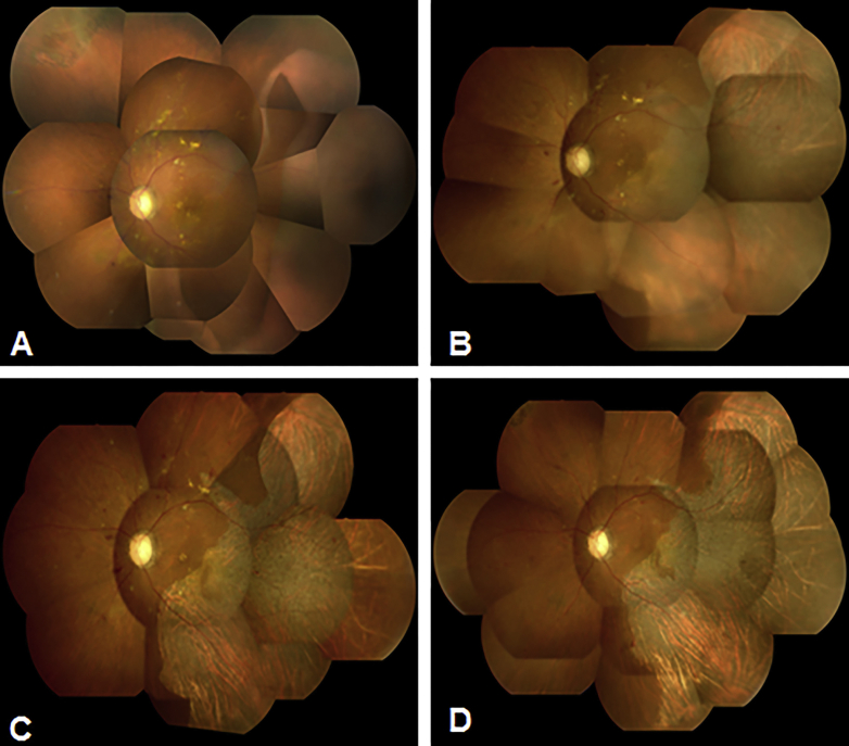
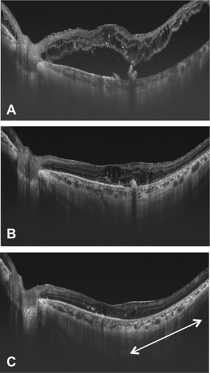
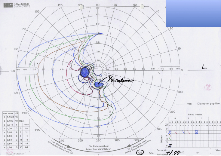
Observations: A 60-year-old man with type 2 diabetes mellitus and end-stage diabetic kidney disease who had gained weight because of fluid overload complained of a visual disturbance in the left eye that had started a few days earlier. The left fundus showed a RPE defect in two temporal quadrants under an extensive serous retinal detachment (SRD) with exacerbation of the original DME. Seven days later, he was admitted for severe edema and pleural effusion. No overt signs of congestive heart failure were noted. On admission, the RPE defect had markedly widened to involve the macula. Spectral-domain optical coherence tomography images showed substantial intraretinal fluid and an extensive SRD with rolled edges of the retinal pigment epithelium, which led to the diagnosis of a RPE tear. The fluid under the SRD was absorbed on the fourth hospital day and the substantial intraretinal fluid resolved on the eleventh day after systemic management of fluid overload only without ophthalmic treatment. The change in the appearance of the RPE area was minimal and the visual field defect remained even after 6 months.
Conclusions and importance: A RPE tear may develop in association with fluid overload in patients with diabetes.
Keywords: Diabetic macular edema; Fluid overload; Retinal pigment epithelial tear; Serous retinal detachment.
Figures
References
-
- Yeo J.H., Marcus S., Murphy R.P. Retinal pigment epithelial tears: patterns and prognosis. Ophthalmology. 1988;95(1):8–13. - PubMed
-
- Gass J.D. Retinal pigment epithelial rip during krypton red laser photocoagulation. Am J Ophthalmol. 1984;98(6):700–706. - PubMed
-
- Dhalla M.S., Blinder K.J., Tewari A., Hariprasad S.M., Apte R.S. Retinal pigment epithelial tear following intravitreal pegaptanib sodium. Am J Ophthalmol. 2006;141(4):752–754. - PubMed
-
- Ishida Y., Kato T., Minamoto A., Yokoyama T., Jian K., Mishima H.K. Retinal pigment epithelial tear in a patient with central serous chorioretinopathy treated with corticosteroids. Retina. 2004;24(4):633–636. - PubMed
Publication types
LinkOut - more resources
Full Text Sources
Other Literature Sources
