

Current Clinical Applications of Diffusion-Tensor Imaging in Neurological Disorders
- PMID: 29504292
- PMCID: PMC5897194
- DOI: 10.3988/jcn.2018.14.2.129
Current Clinical Applications of Diffusion-Tensor Imaging in Neurological Disorders
Abstract
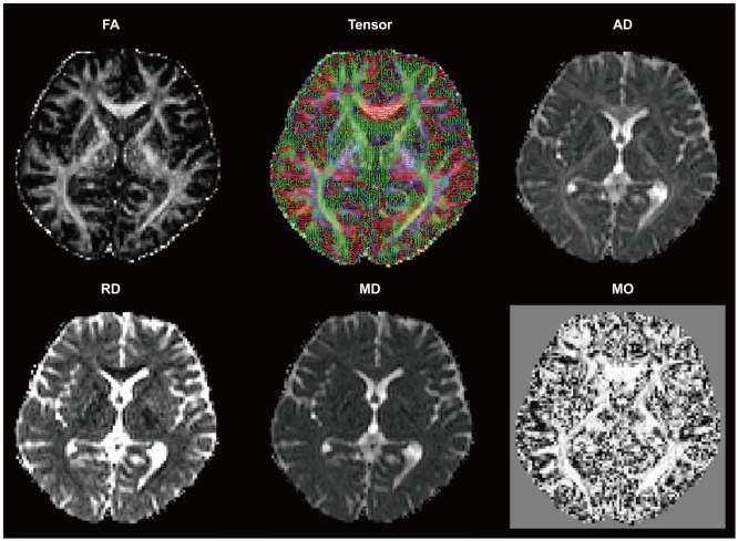
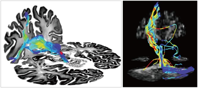
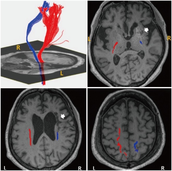
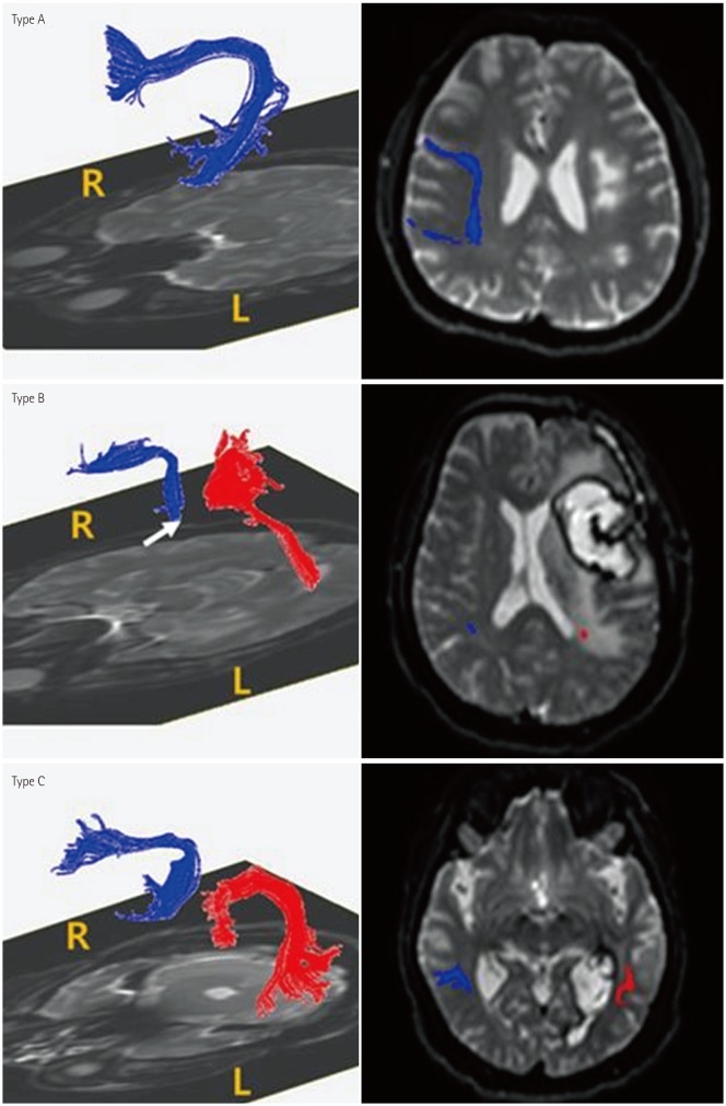
Diffusion-tensor imaging (DTI) is a noninvasive medical imaging tool used to investigate the structure of white matter. The signal contrast in DTI is generated by differences in the Brownian motion of the water molecules in brain tissue. Postprocessed DTI scalars can be used to evaluate changes in the brain tissue caused by disease, disease progression, and treatment responses, which has led to an enormous amount of interest in DTI in clinical research. This review article provides insights into DTI scalars and the biological background of DTI as a relatively new neuroimaging modality. Further, it summarizes the clinical role of DTI in various disease processes such as amyotrophic lateral sclerosis, multiple sclerosis, Parkinson's disease, Alzheimer's dementia, epilepsy, ischemic stroke, stroke with motor or language impairment, traumatic brain injury, spinal cord injury, and depression. Valuable DTI postprocessing tools for clinical research are also introduced.
Keywords: diffusion-tensor imaging; diffusion-tensor imaging scalar; neurological disorders; postprocessing.
Copyright © 2018 Korean Neurological Association.
Conflict of interest statement
The authors have no financial conflicts of interest.
Figures
References
-
- Acosta-Cabronero J, Williams GB, Pengas G, Nestor PJ. Absolute diffusivities define the landscape of white matter degeneration in Alzheimer's disease. Brain. 2010;133:529–539. - PubMed
-
- Abhinav K, Yeh FC, Pathak S, Suski V, Lacomis D, Friedlander RM, et al. Advanced diffusion MRI fiber tracking in neurosurgical and neurodegenerative disorders and neuroanatomical studies: a review. Biochim Biophys Acta. 2014;1842:2286–2297. - PubMed
-
- Dong Q, Welsh RC, Chenevert TL, Carlos RC, Maly-Sundgren P, Gomez-Hassan DM, et al. Clinical applications of diffusion tensor imaging. J Magn Reson Imaging. 2004;19:6–18. - PubMed
-
- Cauley KA, Filippi CG. Diffusion-tensor imaging of small nerve bundles: cranial nerves, peripheral nerves, distal spinal cord, and lumbar nerve roots--clinical applications. AJR Am J Roentgenol. 2013;201:W326–W335. - PubMed
Publication types
LinkOut - more resources
Full Text Sources
Other Literature Sources
Medical
