Clinicopathological features of combined hepatocellular-cholangiocarcinoma with sarcomatous change: Case report and literature review
- PMID: 29504997
- PMCID: PMC5779766
- DOI: 10.1097/MD.0000000000009640
Clinicopathological features of combined hepatocellular-cholangiocarcinoma with sarcomatous change: Case report and literature review
Abstract
Rationale: Combined hepatocellular-cholangiocarcinoma (cHCC-CC) is a rare subtype of primary liver malignancy comprising <1.5% of all primary liver tumors. Sarcomatoid changes in cHCC-CC are even rarer. Due to the rarity of this subtype, its clinicopathological feature is poorly understood. Therefore, here we report 2 tumors.
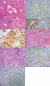
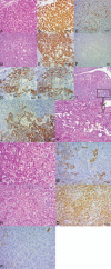
Patient concerns: The first patient was a 44-year-old man with 5-year history of hepatitis B-induced cirrhosis. The resection of right liver revealed a 2.5 × 2.5 × 2 cm tumor mass. Histologically, the tumor showed areas of the typical moderately differentiated HCC. An intermingled adenocarcinoma with pleomorphic and spindle-shaped cells was also identified. The second case involved a 54-year-old man with a history of hepatitis B-induced cirrhosis. A 3.5 × 3 × 3 cm mass was found in the middle left of falciform ligament. Microscopically, the tumor consisted of spindle-shaped sarcomatoid carcinoma cells mixed with typical well-differentiated HCC and well-differentiated CC.
Diagnoses: According to the clinicopathological features, diagnosis of cHCC-CC with sarcomatous change was made.
Interventions: In the first case, right lobectomy of the liver was performed. The second patient underwent laparoscopic, hepatic left lateral lobectomy.
Outcomes: The first patient was alive and well 10 years after the surgical resection without additional treatment. In second case, at 8 months after surgical resection, there was no evidence of recurrence or metastasis.
Lessons: In this report, we describe 2 rare cases of cHCC-CC with sarcomatous change, and findings are helpful for the pathologists would like to further identify the clinicopathological features of this rare tumor.
Copyright © 2017 The Authors. Published by Wolters Kluwer Health, Inc. All rights reserved.
Conflict of interest statement
The authors have no conflicts of interest to disclose.
Figures


References
-
- TohruNakajima, HitoshiKubosawa, YoichiroKondo, et al. Combined hepatocellular-cholangiocarcinoma with variabl sarcomatous transformation. Am J Clin Pathol 1988;90:309. - PubMed
-
- Haratake J, Horie A. An immunohistochemical study of sarcomatoid liver carcinomas. Cancer 1991;68:93–7. - PubMed
-
- Papotti M, Sambataro D, Marchesa P, et al. A combined hepatocellular/cholangiocellular carcinoma with sarcomatoid features. Liver 1997;17:47. - PubMed
-
- Murata M, Miyoshi Y, Iwao K, et al. Combined hepatocellular/cholangiocellular carcinoma with sarcomatoid features: genetic analysis for histogenesis. Hepatol Res 2001;21:220–7. - PubMed
-
- Kim@bullet M-J, Seung K, Lee K, et al. A case of combined hepatocellular and cholangiocarcinoma with neuroendocrine differentiation and sarcomatoid transformation: a case report. Korean J Pathol 2005;39:125–9.
Publication types
MeSH terms
LinkOut - more resources
Full Text Sources
Other Literature Sources
Medical

