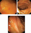
Case report of primary intestinal lymphangiectasia diagnosed in an octogenarian by ileal intubation and by push enteroscopy after missed diagnosis by standard colonoscopy and EGD
- PMID: 29505002
- PMCID: PMC5779771
- DOI: 10.1097/MD.0000000000009649
Case report of primary intestinal lymphangiectasia diagnosed in an octogenarian by ileal intubation and by push enteroscopy after missed diagnosis by standard colonoscopy and EGD
Abstract
Rationale: Primary intestinal lymphangiectasia (PIL) is a rare, presumably congenital lesion that is usually diagnosed in patients < 3 years old, is rarely first diagnosed in adulthood, and when first diagnosed in adulthood typically presents with symptoms for many years. Although PIL is often identified by endoscopic abnormalities, it must be emphasized that the jejunoileum/distal duodenum must be intubated for diagnosis because the lesions are present in these regions. This work demonstrates that 1)-PIL can occur in an octogenarian; 2)-shows that the characteristic endoscopic findings are not found at colonoscopy without terminal ileal intubation; and 3)-may be missed at standard EGD without distal duodenal intubation.
Diagnoses: A patient initially presented at age 83 with symptoms of watery diarrhea, abdominal distention, 5-Kg-weight-gain, and weakness for one month, and had typical clinical findings of PIL including chylous ascites, pleural effusions, bilateral pitting leg edema, hypoalbuminemia, borderline lymphopenia, hypovitaminosis-D, and hypocalcemia. Protein-losing-enteropathy was demonstrated by positive stool tests for alpha-1-antitrypsin. Standard colonoscopy revealed no significant lesions, but terminal ileal intubation during colonoscopy demonstrated creamy-white, punctate, mucosal lesions in terminal ileum, characteristic of lymphangiectasia. EGD with intubation to mid-descending duodenum revealed no significant lesions, but subsequent enteroscopy demonstrated lesions in distal duodenum/proximal jejunum similar to those in terminal ileum characteristic of lymphangiectasia. Histopathologic analysis of lesions of terminal ileum/distal duodenum demonstrated dilated mucosal vessels, confirmed as lymphatic vessels by immunohistochemistry. PIL was diagnosed after excluding secondary causes of intestinal lymphangiectasia.
Interventions/outcomes: Patient placed on standard PIL diet: oral supplements of medium-chain triglycerides, a high protein diet, supplements of fat-soluble vitamins, and avoiding long-chain fatty acids, with marked clinical improvement.
Lessons: This work shows that: 1)-standard EGD and colonoscopy may miss characteristic lesions of PIL, 2)-enteroscopy or terminal ileal intubation at colonoscopy may be required for the diagnosis because lesions are typically located in distal duodenum/jejunoileum; and 3)-PIL can first present in the very elderly even with symptoms of short duration.
Copyright © 2017 The Authors. Published by Wolters Kluwer Health, Inc. All rights reserved.
Conflict of interest statement
The authors have no conflicts of interest to disclose. In particular, MSC, as a consultant of the United States Food and Drug Administration (FDA) Advisory Committee for Gastrointestinal Drugs, affirms that this paper does not discuss any proprietary confidential pharmaceutical data submitted to the FDA. MSC is also a member of the speaker's bureau for AstraZeneca and Daiichi Sankyo, comarketers of Movantik. This work does not discuss any drug manufactured or marketed by AstraZeneca or Daiichi Sankyo.
Figures


Similar articles
-
Primary intestinal lymphangiectasia in an adult patient: A case report and review of literature.World J Gastroenterol. 2020 Dec 28;26(48):7707-7718. doi: 10.3748/wjg.v26.i48.7707. Epub 2020 Dec 8. World J Gastroenterol. 2020. PMID: 33505146 Free PMC article. Review.
-
Primary intestinal lymphangiectasia (Waldmann's disease).Orphanet J Rare Dis. 2008 Feb 22;3:5. doi: 10.1186/1750-1172-3-5. Orphanet J Rare Dis. 2008. PMID: 18294365 Free PMC article. Review.
-
Ileal polypoid lymphangiectasia bleeding diagnosed and treated by double balloon enteroscopy.World J Gastroenterol. 2013 Dec 7;19(45):8440-4. doi: 10.3748/wjg.v19.i45.8440. World J Gastroenterol. 2013. PMID: 24363538 Free PMC article. Review.
-
Primary intestinal lymphangiectasia diagnosed by capsule endoscopy and double balloon enteroscopy.World J Gastrointest Endosc. 2011 Nov 16;3(11):235-40. doi: 10.4253/wjge.v3.i11.235. World J Gastrointest Endosc. 2011. PMID: 22110841 Free PMC article.
-
[Primary intestinal lymphangiectasia (Waldmann's disease)].Rev Med Interne. 2018 Jul;39(7):580-585. doi: 10.1016/j.revmed.2017.07.009. Epub 2017 Sep 1. Rev Med Interne. 2018. PMID: 28867533 Review. French.
Cited by
-
Experience of primary intestinal lymphangiectasia in adults: Twelve case series from a tertiary referral hospital.World J Clin Cases. 2024 Feb 6;12(4):746-757. doi: 10.12998/wjcc.v12.i4.746. World J Clin Cases. 2024. PMID: 38322684 Free PMC article.
-
Primary intestinal lymphangiectasia in an adult patient: A case report and review of literature.World J Gastroenterol. 2020 Dec 28;26(48):7707-7718. doi: 10.3748/wjg.v26.i48.7707. Epub 2020 Dec 8. World J Gastroenterol. 2020. PMID: 33505146 Free PMC article. Review.
-
Small intestinal mucosal abnormalities using video capsule endoscopy in intestinal lymphangiectasia.Orphanet J Rare Dis. 2023 Oct 2;18(1):308. doi: 10.1186/s13023-023-02914-z. Orphanet J Rare Dis. 2023. PMID: 37784188 Free PMC article.
-
Novel Case Report: A Previously Reported, but Pathophysiologically Unexplained, Association Between Collagenous Colitis and Protein-Losing Enteropathy May Be Explained by an Undetected Link with Collagenous Duodenitis.Dig Dis Sci. 2021 Dec;66(12):4557-4564. doi: 10.1007/s10620-020-06804-3. Epub 2021 Feb 4. Dig Dis Sci. 2021. PMID: 33537921 Free PMC article.
-
Elderly Onset Primary Intestinal Lymphangiectasia-A Rare Case.JGH Open. 2025 Jan 22;9(1):e70102. doi: 10.1002/jgh3.70102. eCollection 2025 Jan. JGH Open. 2025. PMID: 39850090 Free PMC article.
References
-
- Wen J, Tang Q, Wu J, et al. Primary intestinal lymphangiectasia: four case reports and a review of the literature. Dig Dis Sci 2010;55:3466–72. - PubMed
-
- Perrault J, Markowitz H. Protein-losing gastroenteropathy and the intestinal clearance of serum-alhpa-1-antitrypsin. Mayo Clin Proc 1984;59:278–9. - PubMed
-
- Hennekam RC, Geerdink RA, Hamel BC, et al. Autosomal recessive intestinal lymphangiectasia and lymphedema with facial anomalies and mental retardation. Am J Med Genet 1989;34:593–600. - PubMed
Publication types
MeSH terms
LinkOut - more resources
Full Text Sources
Other Literature Sources

