. 2018 Jun;154(8):2252-2265.e2.
doi: 10.1053/j.gastro.2018.02.030. Epub 2018 Mar 2.

Neuronal Transforming Growth Factor beta Signaling via SMAD3 Contributes to Pain in Animal Models of Chronic Pancreatitis

Affiliations

Neuronal Transforming Growth Factor beta Signaling via SMAD3 Contributes to Pain in Animal Models of Chronic Pancreatitis

Liansheng Liu et al. Gastroenterology. 2018 Jun.

Abstract

Background & aims: Chronic pancreatitis (CP) is characterized by pancreatic inflammation and fibrosis, associated with increased pancreatic expression of transforming growth factor beta (TGFB). It is not clear how these might contribute to pain. We investigated whether TGFB signaling via SMAD induces sensitization of pancreatic sensory neurons to increase nociception.

Methods: CP was induced in Sprague-Dawley rats by infusion of trinitrobenzene sulfonic acid; some rats were given intrathecal infusions of TGFB1. CP was induced in control mice by administration of cerulein; we also studied β1glo/Ptf1acre-ER mice, which on induction overexpress TGFB1 in pancreatic acinar cells, and TGFBr1f/f-CGRPcreER mice, which have inducible disruption of TGFBr1 in calcitonin gene-related peptide-positive neurons. Dominant negative forms of human TGFBR2 and SMAD3 were overexpressed from viral vectors in rat pancreas. Some rats were given the SMAD3 inhibitors SIS3 or halofuginone. After induction of CP, mice were analyzed for pain in behavior tests or electrophysiologic studies of sensory neurons. Pancreatic nociceptor excitability was examined by patch-clamp techniques and nociception was measured by Von Frey Filament tests for referred somatic hyperalgesia and behavioral responses to pancreatic electrical stimulation. Pancreata were collected from mice and rats and analyzed histologically and by enzyme-linked immunosorbent assay and immunohistochemistry.

Results: Overexpression of TGFB in pancreatic acinar cells of mice and infusion of TGFB1 into rats resulted in sensory neuron hyperexcitability, SMAD3 activation, and increased nociception. This was accompanied by a reduction in the transient A-type current in pancreas-specific sensory neurons in rats, a characteristic of nociceptive sensitization in animal models of CP. Conversely, pancreata from TGFBr1f/f-CGRPcreER mice, rats with pancreatic expression of dominant negative forms of human TGFBR2 or SMAD3, and rats given small molecule inhibitors of SMAD3 had attenuated neuronal sensitization and pain behavior following induction of CP. In contrast to findings from peripheral administration of TGFB1, intrathecal infusion of TGFB1 reduced hyperalgesia in rats with CP.

Conclusions: In pancreata of mice and rats, TGFB promotes peripheral nociceptive sensitization via a direct effect on primary sensory neurons mediated by intra-neuronal SMAD3. This is distinct from the central nervous system, where TGFB reduces nociception. These results provide an explanation for the link between fibrosis and pain in patients with CP. This signaling pathway might be targeted therapeutically to reduce pain in patients with CP.

Keywords: Mouse Model; Neurobiology; Pain Signal Transduction; Pancreas.


Conflict of interest statement

Disclosures: All authors (LL, YZ, MN, QL and PJP) have declared that no conflict of interest exists.

Figures

Figure 1
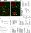
Figure 1. Effect of intra-ductal infusion of TGFB1 on SMAD signaling, pain behavior and excitability of pancreatic sensory neurons
A) SMAD signaling in pancreatic nociceptive neuron. A1: An example of pSmad3 staining (green) in DiI labeled pancreatic neurons (red). Arrow heads indicate DiI labeled pSmad3 positive neurons and arrows indicate DiI labeled pSmad3 negative neurons; A2: Plot showing that pSmad3 signaling was significantly increase in the pancreatic innervated DRG neurons of TGFB-infused rats (*: P<0.001 compared to Vehicle by Student t-test) (n=29-34 DRG sections from 4-5 rats). B) Pancreatic ductal infusion of TGFB1 (1μg, but not 0.1μg) increased pancreatic pain sensitivity (*: P<0.05 compared with control group by Student-Newman-Keuls post-hoc test). Data are presented as mean ± SEM (n=6). C & D) Whole-cell voltage patch-clamp recordings on pancreas-projecting (DiI-positive) DRG neurons. C. Pancreatic ductal infusion of TGFB1 (1μg) increased excitability of these neurons. From left to right: Example of patch-clamp recording, plots of parameters for neural excitability (*: P<0.05 compared to control group by Student T-test). D. Changes in voltage dependent potassium currents in pancreatic neurons. From left to right: Examples of the patch-clamping recording; plots of total inward potassium currents (Itotal); IK currents and IA current (*: P<0.05 compared to control group by Student T-test). Data are presented as mean ± SEM (n=18-39 cells).
Figure 2
Figure 2. Effects of selective overexpression of TGFB1 in the pancreas of β1glo/Ptf1acre-ER mice (induced by tamoxifen) on SMAD signaling and pain behavior in nociceptive neurons
A) SMAD signaling in pancreatic nociceptive neurons was increased in tamoxifen-induced β1glo/Ptf1acre-ER mice. A1. An example of pSmad3 (red) and TRPV1 (green) staining in DRGT8-12 neurons of β1glo/Ptf1acre-ER mice. Arrows indicate TRPV1-positive neurons expressing pSmad3; arrow-heads indicate TRPV1-positive neurons that do not express pSmad3. A2. Plot showing the proportion of TRPV1-positive cells in DRGT8-12 that express pSmad3 was significantly increased in mice after tamoxifen induction (*: P<0.05 compared to corn oil treated group by student t-test). Data are presented as mean ± SEM (n=26-61 sections from 4-6 mice). B) TGFB1 in the pancreas was increased after tamoxifen injection in the transgenic mice as measured by ELISA. (*: P<0.05 compared to corn oil group by student t-test). Data are presented as mean ± SEM (n=6-7 mice). C) Referred somatic sensitivity, as measured by VFF testing, was increased after TGFB1 over-expression induced by tamoxifen (*: P <0.05 compared with corn oil treated group by Student-Newman-Keuls post hoc test). Data are presented as mean ± SEM (n=6-7 mice).
Figure 3
Figure 3. Effects of downregulation of TGFBRI on SMAD signaling in nociceptive neurons and pain behavior in TGFBRIf/f-CGRPcreER mice with cerulein-induced CP
A1) An example of pSmad3 (red) and TRPV1 (green) staining in the DRGT9-10 of TGFBRIf/f-CGRPcreER mice. The arrows indicated same as described in Figure2. A2) Plot showing reduction of pSmad3 in TRPV1-positive DRG neurons after tamoxifen induction in both CP and control groups (*: P <0.05 compared to corn oil treated group in same model by Student-Newman-Keuls post hoc test). B) Tamoxifen-induced downregulation of TGFBRI attenuated the increased pain sensitivity in mice with CP (* P <0.05 for comparison with corn oil/cerulein group; #: P<0.05 for comparison with Corn oil/saline group at same VFF strength by Student-Newman-Keuls post hoc test). Data were presented as mean ± SEM (n=6-7 mice). C) In vitro TGFB1 on DRG neuronal activity in TGFBRI knockout mice. TGFBRI knockout was induced by administration of tamoxifen for 5 days. DRG neurons from the control or TGFBRI knockout mice were incubated with 10nM TGFB1 for 48 hours and electrophysiological responses were examined by whole cell voltage patch clamping. * P <0.05 for comparison with vehicle group treated with corn oil; #: P<0.05 for comparison with TGFB1 incubated cells in corn oil treated mice by Student-Newman-Keuls post hoc test). Data were presented as mean ± SEM (n=10-12 cells)
Figure 4
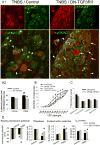
Figure 4. Effects of knock-down of TGFBRII by DN-TGFBRII-HSV on SMAD signaling, pancreatic neuronal excitability and pain behavior in CP rats
A) SMAD signaling in pancreatic nociceptive neuron. A1: An example of pSmad3 staining (green) in DiI labeled pancreatic neurons (red). Arrows indicated as described in Figure1; A2: Plot showing that CP-induced increase in pSmad3 signaling was significantly reduced in DN-TGFBRII-HSV injected rats (*: P<0.05 compared to TNBS/Control group; #: P< 0.05 compared to Saline/Control group by Student-Newman-Keuls post hoc test) (n=27-48 DRG sections from 4-5 rats). B): CP-induced hyperalgesia was significantly reduced in rats that received DN-TGFBRII-HSV. * P<0.05 for comparison with TNBS/control group; #: P<0.05 for comparison with vehicle/control group at same VFF strength by Student-Newman-Keuls post hoc test (n=5 rats). C): Pathological scores CP were not reversed by treatment with DN-TGFBRII-HSV. None of the parameters was significantly different between control- and DN-TGFBRII-HSV treated CP rats. Data were present as mean ± SEM (n = 4-5 rats). D): DN-TGFBRII-HSV injection prevented the increase in the excitability of pancreatic neurons in DRGT8-12 of rats with TNBS-induced CP (*: P<0.05 compared to control virus treated CP rats, #: P<0.05 comparing to vehicle rats with same viral treatment by Student-Newman-Keuls post hoc test) (n=23-39 cells).
Figure 5
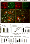
Figure 5. Effects of knock-down of SMAD3 by DN-SMAD3-HSV on SMAD signaling, pancreatic neuronal excitability, pain behavior and histology of pancreas in TNBS-CP rats
A): SMAD signaling in pancreatic neurons. A1: An example of pSmad3 staining (green) in Di-I labeled pancreatic neurons (red). Arrows indicated as described in Figure1; A2: Plot showing that CP-induced increase in pSmad3 was significantly reduced in DN-Smad3-HSV injected rats. (*: P<0.05 compared to Control virus group in the same group; #: P< 0.05 compared to Saline/Control group by Student-Newman-Keuls post hoc test) (n=48-58 DRG sections from 4-5 rats). B): CP-induced hyperalgesia was significantly reduced in rats received DN-Smad3-HSV as measured by VFF responses (* P <0.05 for comparison with TNBS/control group; #: P<0.05 for comparison with vehicle/control group at same VFF strength by Student-Newman-Keuls post hoc test (n=5 rats). C): Pathological scores of CP were not prevented by treatment with DN-SMAD3-HSV. None of the parameters was significantly different between control- and DN-SMAD3-HSV treated CP rats. Data were present as mean ± SEM (n = 4-5 rats) D): DN-Smad3-HSV injection reversed CP-induced changes in excitability of pancreatic-innervated neurons (*: P<0.05 compared to control virus treated CP rats, #: P<0.05 comparing to vehicle rats with same viral treatment by Student-Newman-Keuls post hoc test) (n=40-55 cells in 5 rats).
Figure 6
Figure 6. Effects of SIS3 (A) and halofuginone (B), two small molecule SMAD3 inhibitors, on TGFB-induced sensory neuronal excitability in vitro, and SMAD signaling and pain behavior in CP-rats
A-1 and A-2: Whole-cell voltage recordings of DRG neurons culture exposed to exogenous TGFB1 (10 ng/ml) with and without SIS3 (0.3μM) for 2 days. A-1: Left to right: Example of evoked action potentials after incubation with TGFB; plot of resting membrane potential, rheobase and evoked action potentials. A-2: Left to right: Examples of total inward potassium currents, IK currents and IA currents in sensory neuron and plots of the same. SIS3 prevented TGFB1-induced reduction of IA current (*: P<0.05 comparing to TGFB1/vehicle group, #: P<0.05, compared to control/vehicle group by Student-Newman-Keuls post hoc test). (n=9-17 cells). A-3: SMAD signaling in vivo. Left panel: an example of pSmad3 staining (green) in DiI labeled pancreatic neurons (zoomed-in images in bottom row). Arrows indicated same as described in Figure1. Right panel shows the plot of analysis for pSmad3 staining (*: P<0.05 comparing to vehicle treated TNBS group, #: P<0.05 comparing to control/vehicle group) (n=45-58 DRG sections). A-4 and A-5: Treatment with SIS3 (2.5 mg/kg, i.p.) for 7 days attenuated responses to VFF (A-4) and electric stimulation (ES, A-5) in CP-rats (*: P<0.05 compared to CP/vehicle rats, #: P<0.05 compared to control/vehicle rats) (n=7-8 rats). B) Effects of halofuginone B-1: Western blot showed that pSMAD3 expression was significantly reduced in the DRGs from both control and CP rats (* P<0.05, comparing to the vehicle treated rats with same pre-treatment). B-2 and B-3: Treatment with halofuginone for 14 days attenuated responses to VFF (B-2) and electric stimulation (B-3) in CP-rats (*: P<0.05 compared to CP/vehicle rats, #: P<0.05 compared to control/vehicle rats, by Student-Newman-Keuls post hoc test). Data were presented as mean ± SEM (n=7-8).
Figure 7
Figure 7. Intrathecal infusion of TGFB1 suppressed hyperalgesia in CP rats as examined by VFF (A) and electrode stimulation (B)
Intrathecal infusion of TGFB1 (5μg/rat) into T9-10 level of CP rats for 2 weeks significantly reduced CP-induced hyperalgesia in both VFF and ES tests (*: P<0.05 compared to CP/vehicle rats, #: P<0.05 compared to control/vehicle rats, by Student-Newman-Keuls post hoc test). Data were presented as Mean ± SEM (n=5-6).

Similar articles

Cited by

References

    1. Mullady DK, Yadav D, Amann ST, et al. Type of pain, pain-associated complications, quality of life, disability and resource utilisation in chronic pancreatitis: a prospective cohort study. Gut. 2011;60:77–84. - PMC - PubMed
    1. Mokrowiecka A, Pinkowski D, Malecka-Panas E. Assessment of quality of life in patients with chronic pancreatitis. Med Sci Monit. 2011;17:CR583–8. - PMC - PubMed
    1. Amann ST, Yadav D, Barmada MM, et al. Physical and mental quality of life in chronic pancreatitis: a case-control study from the North American Pancreatitis Study 2 cohort. Pancreas. 2013;42:293–300. - PMC - PubMed
    1. Crosby J, Bellin MD, Radosevich DM, et al. Gastrointestinal symptoms before and after total pancreatectomy with islet autotransplantation: the role of pancreatic enzyme dosing and adherence. Pancreas. 2015;44:453–8. - PMC - PubMed
    1. Bellin MD, Gelrud A, Arreaza-Rubin G, et al. Total pancreatectomy with islet autotransplantation: summary of an NIDDK workshop. Ann Surg. 2015;261:21–9. - PMC - PubMed

Publication types

MeSH terms

Substances