. 2018 Apr;48(5):359-369.
doi: 10.1016/j.ijpara.2018.01.002. Epub 2018 Mar 2.

Heligmosomoides polygyrus Venom Allergen-like Protein-4 (HpVAL-4) is a sterol binding protein

Affiliations

Heligmosomoides polygyrus Venom Allergen-like Protein-4 (HpVAL-4) is a sterol binding protein

Oluwatoyin A Asojo et al. Int J Parasitol. 2018 Apr.

Abstract

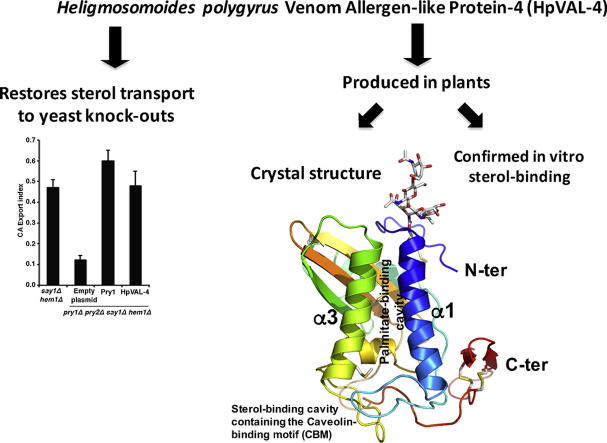
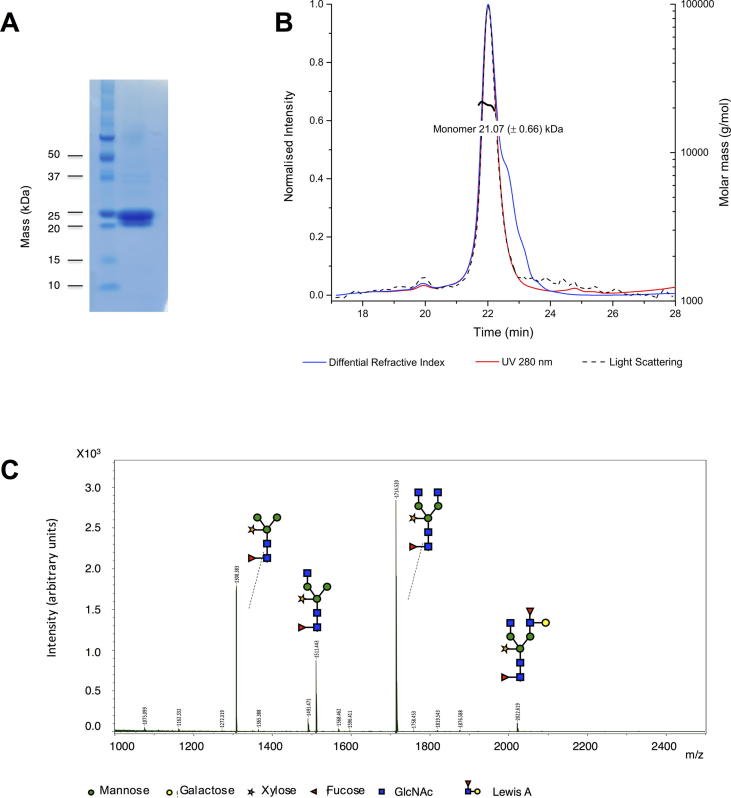
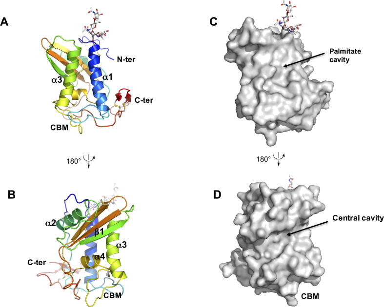
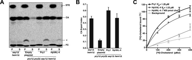
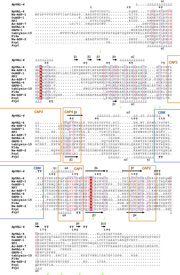
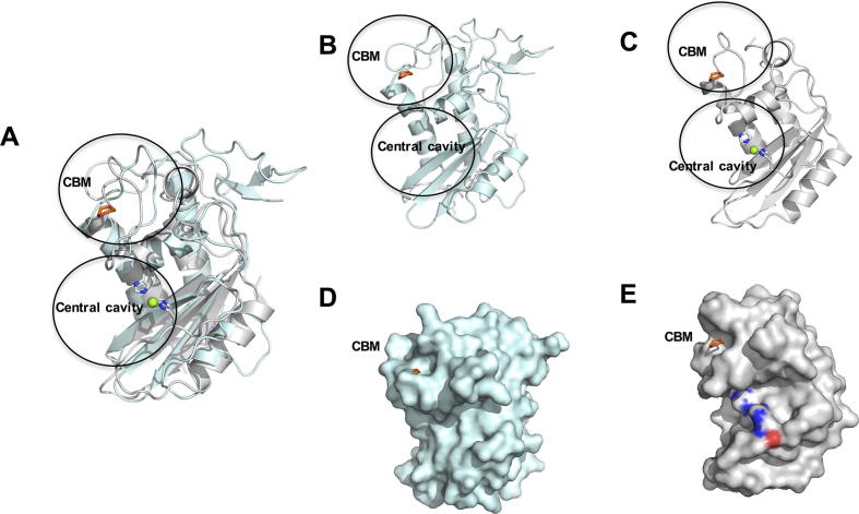
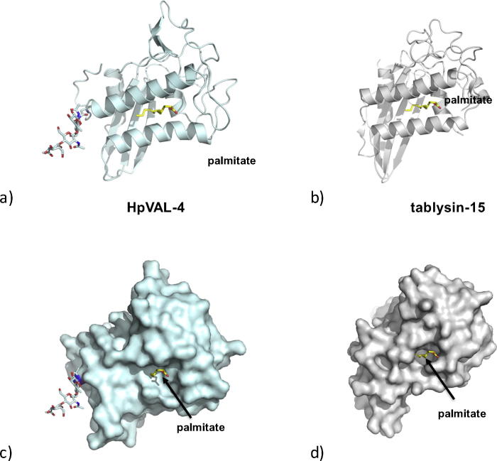
Heligmosomoides polygyrus bakeri is a model parasitic hookworm used to study animal and human helminth diseases. During infection, the parasite releases excretory/secretory products that modulate the immune system of the host. The most abundant protein family in excretory/secretory products comprises the venom allergen-like proteins (VALs), which are members of the SCP/TAPS (sperm-coating protein/Tpx/antigen 5/pathogenesis related-1/Sc7) superfamily. There are >30 secreted Heligmosomoides polygyrus VAL proteins (HpVALs) and these proteins are characterised by having either one or two 15 kDa CAP (cysteine-rich secretory protein (CRISP)/antigen 5/pathogenesis related-1) domains. The first known HpVAL structure, HpVAL-4, refined to 1.9 Å is reported. HpVAL-4 was produced as a homogeneously glycosylated protein in leaves of Nicotiana benthamiana infiltrated with recombinant plasmids, making this plant expression platform amenable for the production of biological products. The overall topology of HpVAL-4 is a three layered αβα sandwich between a short N-terminal loop and a C-terminal cysteine rich extension. The C-terminal cysteine rich extension has two strands stabilized by two disulfide bonds and superposes well with the previously reported extension from the human hookworm Necator americanus Ancylostoma secreted protein-2 (Na-ASP-2). The N-terminal loop is connected to alpha helix 2 via a disulfide bond previously observed in Na-ASP-2. HpVAL-4 has a central cavity that is more similar to the N-terminal CAP domain of the two CAP Na-ASP-1 from Necator americanus. Unlike Na-ASP-2, mammalian CRISP, and the C-terminal CAP domain of Na-ASP-1, the large central cavity of HpVAL-4 lacks the two histidines required to coordinate divalent cations. HpVAL-4 has both palmitate-binding and sterol-binding cavities and is able to complement the in vivo sterol export phenotype of yeast mutants lacking their endogenous CAP proteins. More studies are required to determine endogenous binding partners of HpVAL-4 and unravel the possible impact of sterol binding on immune-modulatory functions.

Keywords: Cysteine-rich secretory protein (CRISP); Excretory–secretory products; Pathogenesis related-1 (PR-1); Sperm coating protein (SCP); Sterol binding; Testis specific proteins (Tpx); Venom antigen 5.


Figures

None
Graphical abstract
Fig. 1
Fig. 1
Protein purity and characterisation of Heligmosomoides polygyrus Venom Allergen-like Protein-4 (HpVAL-4). (A) Coomasie-stained SDS gel reveals the purity of recombinant HpVAL-4 and its monomeric mass of ∼22 kDa. (B) Size exclusion chromatography multi-angle light scattering analysis reveals that HpVAL-4 is an ∼21 kDa monomer in solution. (C) N-glycan composition of plant-produced HpVAL-4.
Fig. 2
Fig. 2
Crystal structure of Heligmosomoides polygyrus Venom Allergen-like Protein-4 (HpVAL-4). (A) Cartoon of a monomer of HpVAL-4 rainbow colours from amino (blue) to carboxyl terminus (red). The two longest helices α1 and α3 that form the palmitate cavity and the caveolin binding motif loop are indicated, while glycans and disulfide bridges are shown in stick form (coloured by elements: blue for N, white for C, red for O, and yellow for S). (B) Rotation (180 degrees) of the monomer allows better visualisation of the strand (β1) and helix (α4) that form the central cavity. (C, D) Surface representations of the views for A and B, respectively.
Fig. 3
Fig. 3
Heligmosomoides polygyrus Venom Allergen-like Protein-4 (HpVAL-4) exports and binds cholesterol in vivo and in vitro. (A) Expression of HpVAL-4 complements the sterol export defect of yeast cells lacking their endogenous cysteine-rich secretory proteins, antigen 5, and pathogenesis-related 1 (CAP) proteins (Pry1 and Pry2). Heme-deficient cells of the indicated genotype containing either an empty plasmid or a plasmid with Pry1 or HpVAL-4 were radiolabeled with [14C]cholesterol overnight, washed and diluted in fresh media to allow for export of cholesterol and cholesteryl acetate. Lipids were extracted from the cell pellet (P) and the culture supernatant (S), and separated by thin layer chromatography. The positions of free cholesterol (FC), cholesteryl acetate (CA) and steryl esters (STE) are indicated. The star marks the position of an unidentified cholesterol derivative. (B) Quantification of the export of cholesteryl acetate in yeast cells lacking their endogenous CAP proteins when complemented with empty plasmid, Pry1 or HpVAL-4. The export index indicates the relative percentages of cholesteryl acetate that is exported by the cells (ratio between the extracellular cholesteryl acetate and the sum of intra- and extra-cellular cholesteryl acetate). Data represent mean ± S.D. of two independent experiments. (C) HpVAL-4 binds cholesterol in vitro. Purified HpVAL-4 protein (100 pmol) was incubated with the indicated concentration of [3H]-cholesterol in the presence (HpVAL-4 + 400 pmol cholesterol) or absence of unlabeled competitor ligand (HpVAL-4). The previously determined Pry1 activity is also shown.
Fig. 4
Fig. 4
Comparison of Heligmosomoides polygyrus Venom Allergen-like Protein-4 (HpVAL-4) with selected members of its superfamily. The sequences were aligned with clustalW2 and the secondary structural features were illustrated with the coordinates of HpVAL-4 and Pry1 using ESPript (Gouet et al., 2003). The different secondary structure elements shown are alpha helices (α), 310-helices (η), beta strands (β), and beta turns (TT). Identical residues are shown in red shading, and conserved residues are in red text. The locations of the cysteine residues involved in disulfide bonds are numbered in green. The location of the caveolin binding motif loop is shown in blue bars and the signature cysteine-rich secretory protein (CRISP) motifs are identified with orange bars. The representative structures are Na-ASP-2 (Asojo et al., 2005), Pry1 (Darwiche et al., 2016), Na-ASP-1 (Asojo, 2011), tablysin-15 (Ma et al., 2011), Golgi-Associated plant Pathogenesis Related protein-1, GAPR-1 (van Galen et al., 2012), Ostertagia ostertagi activation-associated secreted protein-1, OoASP-1 (Borloo et al., 2013), Ancylostoma caninum Ancylostoma secreted protein-7, Ac-ASP-7 (Osman et al., 2012), Schistosoma mansoni Venom Allergen-like Protein-4, SmVAL4, (Kelleher et al., 2015), and Solanum lycopersicum pathogenesis-related protein, P14A (Fernandez et al., 1997).
Fig. 5
Fig. 5
Comparison of Heligmosomoides polygyrus Venom Allergen-like Protein-4 (HpVAL-4) with pathogen-related yeast protein 1 (Pry1). (A) The superposed ribbon structure of HpVAL-4 (cyan) and Pry1 (grey) reveals the conformational flexibility of the caveolin binding motif which contains the 1,2-dioxane from the Pry1 structure (shown in red). The central histidines that coordinate cations in Pry1 are coloured by elements with blue for N, white for C, red for O. Mg2+ is shown as a green sphere. Ribbon diagrams of the same view of (B) HpVAL-4 and (C) Pry1. The sizes of the cavities are evident from the surface plot of the same view of (D) HpVAL-4 and (E) Pry1.
Fig. 6
Fig. 6
The palmitate binding cavity. (A) Ribbon diagram of the putative palmitate-binding cavity of Heligmosomoides polygyrus Venom Allergen-like Protein-4 (HpVAL-4) based on (B) the palmitate binding of tablysin-15. Surface representations of same view of the palmitate binding cavity of (C) HpVAL-4 and (D) tablysin-15, with palmitate shown as yellow sticks and the glycosylation site on HpVAL-4 as other coloured sticks.

Similar articles

Cited by

References

    1. Adams P.D., Afonine P.V., Bunkoczi G., Chen V.B., Davis I.W., Echols N., Headd J.J., Hung L.W., Kapral G.J., Grosse-Kunstleve R.W., McCoy A.J., Moriarty N.W., Oeffner R., Read R.J., Richardson D.C., Richardson J.S., Terwilliger T.C., Zwart P.H. PHENIX: a comprehensive Python-based system for macromolecular structure solution. Acta Crystallogr. D. 2010;66:213–221. - PMC - PubMed
    1. Asojo O.A. Structure of a two-CAP-domain protein from the human hookworm parasite Necator americanus. Acta Crystallogr. D. 2011;67:455–462. - PMC - PubMed
    1. Asojo O.A., Goud G., Dhar K., Loukas A., Zhan B., Deumic V., Liu S., Borgstahl G.E., Hotez P.J. X-ray structure of Na-ASP-2, a pathogenesis-related-1 protein from the nematode parasite, Necator americanus, and a vaccine antigen for human hookworm infection. J. Mol. Biol. 2005;346:801–814. - PubMed
    1. Asojo O.A., Koski R.A., Bonafe N. Structural studies of human glioma pathogenesis-related protein 1. Acta Crystallogr. D. 2011;67:847–855. - PMC - PubMed
    1. Baroni R.M., Luo Z., Darwiche R., Hudspeth E.M., Schneiter R., Pereira G.A.G., Mondego J.M.C., Asojo O.A. Crystal Structure of MpPR-1i, a SCP/TAPS protein from Moniliophthora perniciosa, the fungus that causes Witches' Broom Disease of Cacao. Sci. Rep. 2017;7:7818. - PMC - PubMed

Publication types