

Selective cytotoxicity of the herbal substance acteoside against tumor cells and its mechanistic insights
- PMID: 29505920
- PMCID: PMC5952579
- DOI: 10.1016/j.redox.2018.02.015
Selective cytotoxicity of the herbal substance acteoside against tumor cells and its mechanistic insights
Abstract
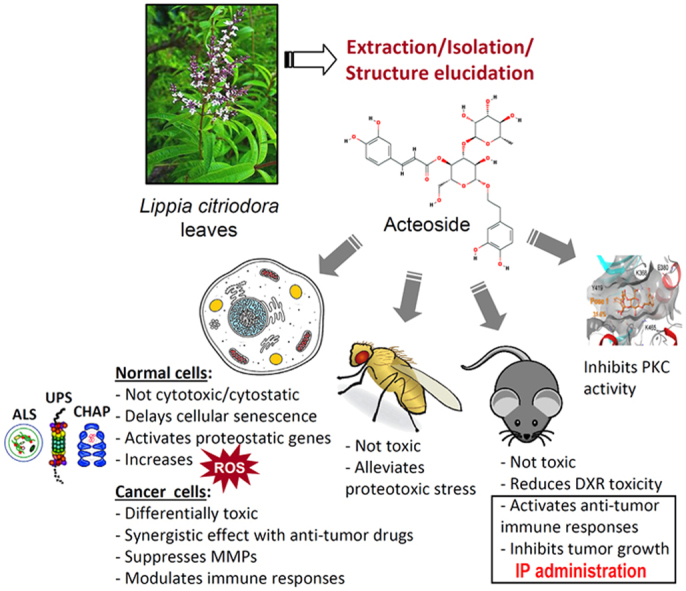
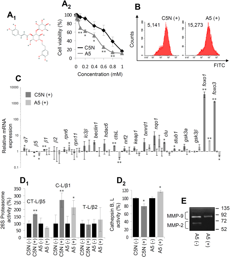
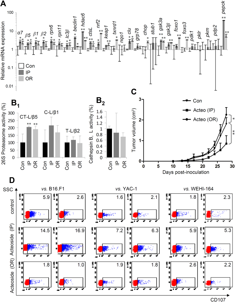
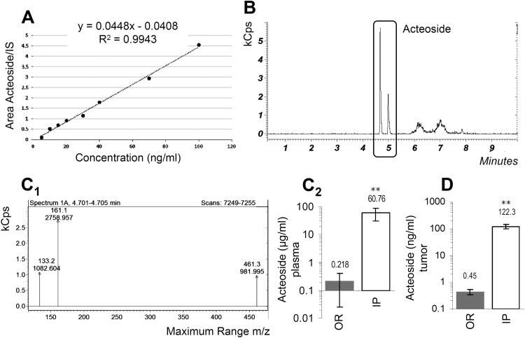
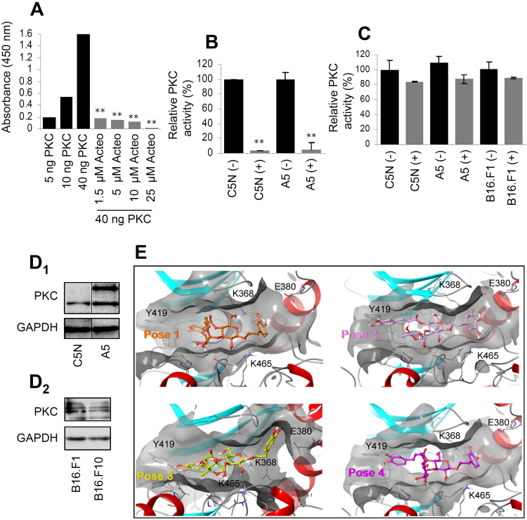
Natural products are characterized by extreme structural diversity and thus they offer a unique source for the identification of novel anti-tumor agents. Herein, we report that the herbal substance acteoside being isolated by advanced phytochemical methods from Lippia citriodora leaves showed enhanced cytotoxicity against metastatic tumor cells; acted in synergy with various cytotoxic agents and it sensitized chemoresistant cancer cells. Acteoside was not toxic in physiological cellular contexts, while it increased oxidative load, affected the activity of proteostatic modules and suppressed matrix metalloproteinases in tumor cell lines. Intraperitoneal or oral (via drinking water) administration of acteoside in a melanoma mouse model upregulated antioxidant responses in the tumors; yet, only intraperitoneal delivery suppressed tumor growth and induced anti-tumor-reactive immune responses. Mass-spectrometry identification/quantitation analyses revealed that intraperitoneal delivery of acteoside resulted in significantly higher, vs. oral administration, concentration of the compound in the plasma and tumors of treated mice, suggesting that its in vivo anti-tumor effect depends on the route of administration and the achieved concentration in the tumor. Finally, molecular modeling studies and enzymatic activity assays showed that acteoside inhibits protein kinase C. Conclusively, acteoside holds promise as a chemical scaffold for the development of novel anti-tumor agents.
Keywords: Acteoside; Cancer; Immunomodulation; Natural compound; Oxidative stress; Proteostasis.
Copyright © 2018 The Authors. Published by Elsevier B.V. All rights reserved.
Figures
Similar articles
-
Inhibitory activity of acteoside in melanoma via regulation of the ERβ-Ras/Raf1-STAT3 pathway.Arch Biochem Biophys. 2021 Oct 15;710:108978. doi: 10.1016/j.abb.2021.108978. Epub 2021 Jun 23. Arch Biochem Biophys. 2021. PMID: 34174222
-
Antimetastatic activity of acteoside, a phenylethanoid glycoside.Biol Pharm Bull. 2002 May;25(5):666-8. doi: 10.1248/bpb.25.666. Biol Pharm Bull. 2002. PMID: 12033512
-
Acteoside inhibits alpha-MSH-induced melanin production in B16 melanoma cells by inactivation of adenyl cyclase.J Pharm Pharmacol. 2009 Oct;61(10):1347-51. doi: 10.1211/jpp/61.10.0011. J Pharm Pharmacol. 2009. PMID: 19814867
-
Anticancer effects of acteoside: Mechanistic insights and therapeutic status.Eur J Pharmacol. 2022 Feb 5;916:174699. doi: 10.1016/j.ejphar.2021.174699. Epub 2021 Dec 14. Eur J Pharmacol. 2022. PMID: 34919888 Review.
-
Advanced research on acteoside for chemistry and bioactivities.J Asian Nat Prod Res. 2011 May;13(5):449-64. doi: 10.1080/10286020.2011.568940. J Asian Nat Prod Res. 2011. PMID: 21534045 Review.
Cited by
-
A Survey of Naturally Occurring Molecules as New Endoplasmic Reticulum Stress Activators with Selective Anticancer Activity.Cancers (Basel). 2022 Dec 31;15(1):293. doi: 10.3390/cancers15010293. Cancers (Basel). 2022. PMID: 36612288 Free PMC article.
-
The pharmacokinetic property and pharmacological activity of acteoside: A review.Biomed Pharmacother. 2022 Sep;153:113296. doi: 10.1016/j.biopha.2022.113296. Epub 2022 Jun 17. Biomed Pharmacother. 2022. PMID: 35724511 Free PMC article. Review.
-
Antitumor Potential of Lippia citriodora Essential Oil in Breast Tumor-Bearing Mice.Antioxidants (Basel). 2021 May 30;10(6):875. doi: 10.3390/antiox10060875. Antioxidants (Basel). 2021. PMID: 34070804 Free PMC article.
-
Verbascoside: comprehensive review of a phenylethanoid macromolecule and its journey from nature to bench.Inflammopharmacology. 2024 Oct;32(5):2729-2751. doi: 10.1007/s10787-024-01555-3. Epub 2024 Aug 20. Inflammopharmacology. 2024. PMID: 39162902 Review.
-
Analysis of Centranthera grandiflora Benth Transcriptome Explores Genes of Catalpol, Acteoside and Azafrin Biosynthesis.Int J Mol Sci. 2019 Nov 29;20(23):6034. doi: 10.3390/ijms20236034. Int J Mol Sci. 2019. PMID: 31795510 Free PMC article.
References
-
- Hanahan D., Weinberg R. Hallmarks of cancer: the next generation. Cell. 2011;144:646–674. - PubMed
-
- Trougakos I., Sesti F., Tsakiri E., Gorgoulis V. Non-enzymatic post-translational protein modifications and proteostasis network deregulation in carcinogenesis. J. Proteom. 2013;92:274–298. - PubMed
-
- Höhn A., Jung T., Grune T. Pathophysiological importance of aggregated damaged proteins. Free Radic. Biol. Med. 2014;71:70–89. - PubMed
Publication types
MeSH terms
Substances
LinkOut - more resources
Full Text Sources
Other Literature Sources
