

Pigmentary mosaicism: a review of original literature and recommendations for future handling
- PMID: 29506540
- PMCID: PMC5839061
- DOI: 10.1186/s13023-018-0778-6
Pigmentary mosaicism: a review of original literature and recommendations for future handling
Abstract
Background: Pigmentary mosaicism is a term that describes varied patterns of pigmentation in the skin caused by genetic heterogeneity of the skin cells. In a substantial number of cases, pigmentary mosaicism is observed alongside extracutaneous abnormalities typically involving the central nervous system and the musculoskeletal system. We have compiled information on previous cases of pigmentary mosaicism aiming to optimize the handling of patients with this condition. Our study is based on a database search in PubMed containing papers written in English, published between January 1985 and April 2017. The search yielded 174 relevant and original articles, detailing a total number of 651 patients.
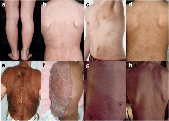
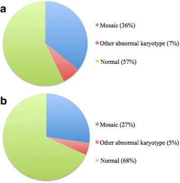
Results: Forty-three percent of the patients exhibited hyperpigmentation, 50% exhibited hypopigmentation, and 7% exhibited a combination of hyperpigmentation and hypopigmentation. Fifty-six percent exhibited extracutaneous manifestations. The presence of extracutaneous manifestations in each subgroup varied: 32% in patients with hyperpigmentation, 73% in patients with hypopigmentation, and 83% in patients with combined hyperpigmentation and hypopigmentation. Cytogenetic analyses were performed in 40% of the patients: peripheral blood lymphocytes were analysed in 48%, skin fibroblasts in 5%, and both analyses were performed in 40%. In the remaining 7% the analysed cell type was not specified. Forty-two percent of the tested patients exhibited an abnormal karyotype; 84% of those presented a mosaic state and 16% presented a non-mosaic structural or numerical abnormality. In patients with extracutaneous manifestations, 43% of the cytogenetically tested patients exhibited an abnormal karyotype. In patients without extracutaneous manifestations, 32% of the cytogenetically tested patients exhibited an abnormal karyotype.
Conclusion: We recommend a uniform parlance when describing the clinical picture of pigmentary mosaicism. Based on the results found in this review, we recommend that patients with pigmentary mosaicism undergo physical examination, highlighting with Wood's light, and karyotyping from peripheral blood lymphocytes and skin fibroblasts. It is important that both patients with and without extracutaneous manifestations are tested cytogenetically, as the frequency of abnormal karyotype in the two groups seems comparable. According to the results only a minor part of patients, especially those without extracutaneous manifestations, are tested today reflecting a need for change in clinical practice.
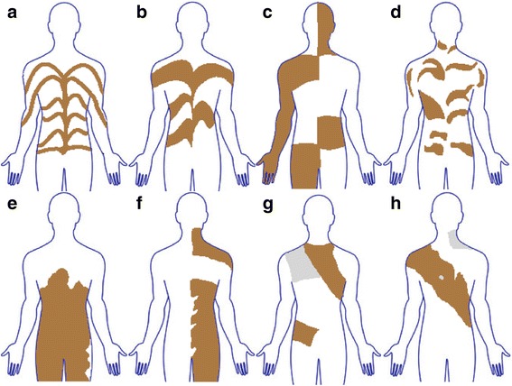
Keywords: Blaschko’s lines; Hyperpigmentation; Hypomelanosis of Ito; Hypopigmentation; Linear and whorled nevoid hypermelanosis; Pigmentary mosaicism.
Conflict of interest statement
Ethics approval and consent to participate
Not applicable.
Consent for publication
Not applicable.
Competing interests
The authors declare that they have no competing interests.
Publisher’s Note
Springer Nature remains neutral with regard to jurisdictional claims in published maps and institutional affiliations.
Figures
References
-
- Happle R. Mosaicism in human skin. 1. Berlin: Springer; 2014.
Publication types
MeSH terms
LinkOut - more resources
Full Text Sources
Other Literature Sources
Miscellaneous
