. 2018 Mar 5;17(1):102.
doi: 10.1186/s12936-018-2251-3.

Experimental malaria-associated acute respiratory distress syndrome is dependent on the parasite-host combination and coincides with normocyte invasion

Affiliations

Experimental malaria-associated acute respiratory distress syndrome is dependent on the parasite-host combination and coincides with normocyte invasion

Leen Vandermosten et al. Malar J. .

Abstract

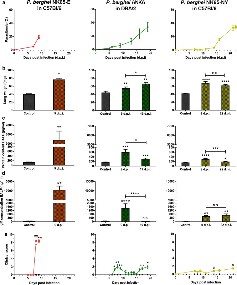
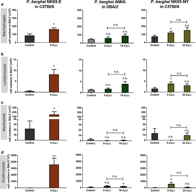
Background: Malaria-associated acute respiratory distress syndrome (MA-ARDS) is a complication of malaria with a lethality rate of up to 80% despite anti-malarial treatment. It is characterized by a vast infiltration of leukocytes, microhaemorrhages and vasogenic oedema in the lungs. Previously, a mouse model for MA-ARDS was developed by infection of C57BL/6 mice with the Edinburgh line NK65-E of Plasmodium berghei.

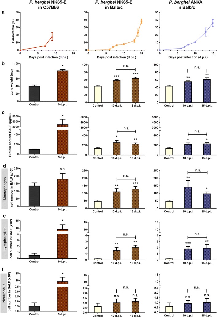
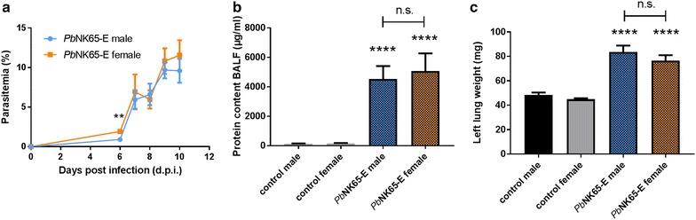
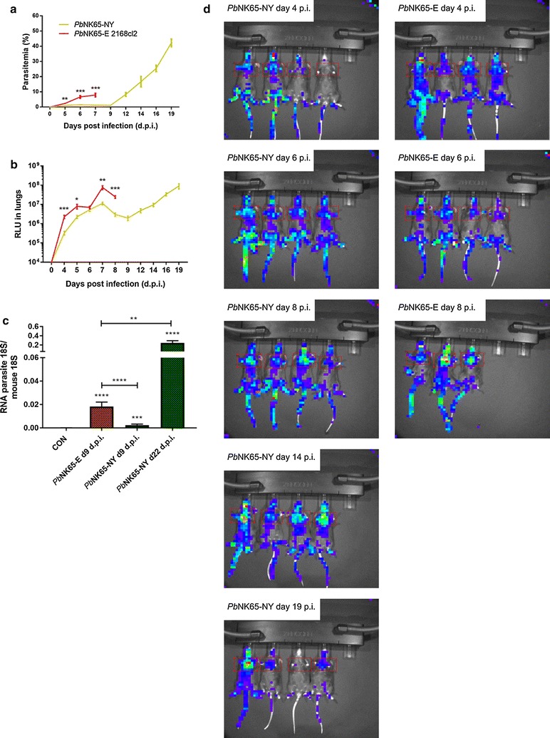
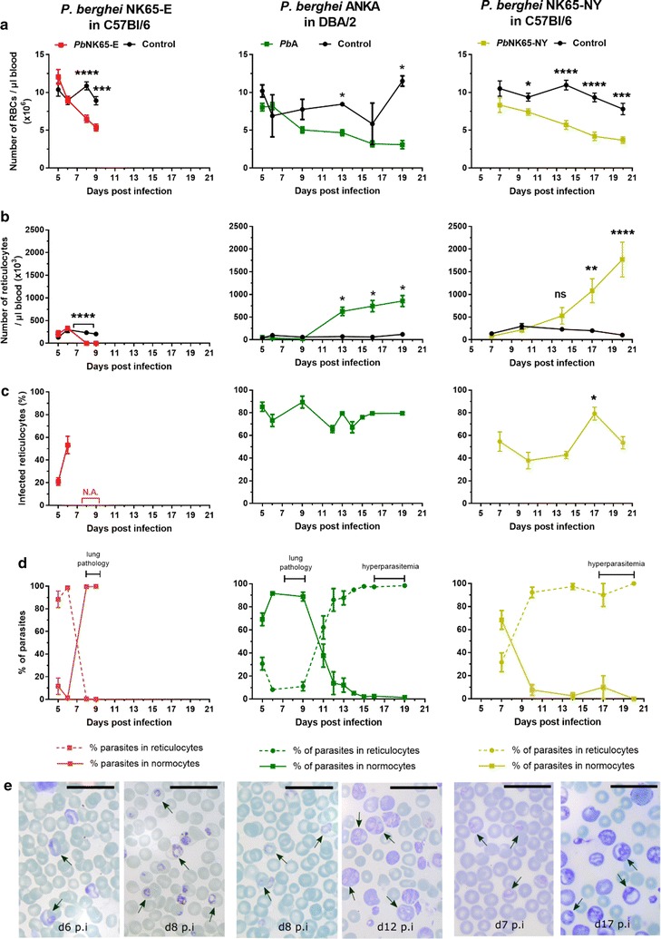
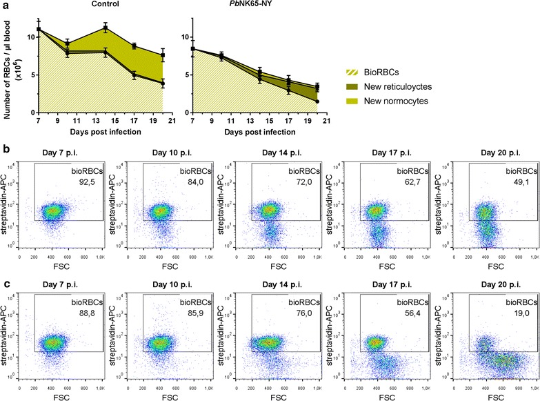
Results: Here, both host and parasite factors were demonstrated to play crucial roles in the development and severity of lung pathology. In particular, the genetic constitution of the host was an important determinant in the development of MA-ARDS. Both male and female C57BL/6, but not BALB/c, mice developed MA-ARDS when infected with P. berghei NK65-E. However, the New York line of P. berghei NK65 (NK65-NY) did not induce demonstrable MA-ARDS, despite its accumulation in the lungs and fat tissue to a similar or even higher extent as P. berghei NK65-E. These two commonly used lines of P. berghei differ in their red blood cell preference. P. berghei NK65-NY showed a stronger predilection for reticulocytes than P. berghei NK65-E and this appeared to be associated with a lower pathogenicity in the lungs. The pulmonary pathology in the C57BL/6/P. berghei NK65-E model was more pronounced than in the model with infection of DBA/2 mice with P. berghei strain ANKA. The transient lung pathology in DBA/2 mice infected with P. berghei ANKA coincided with the infection phase in which parasites mainly infected normocytes. This phase was followed by a less pathogenic phase in which P. berghei ANKA mainly infected reticulocytes.

Conclusions: The propensity of mice to develop MA-ARDS during P. berghei infection depends on both host and parasite factors and appears to correlate with RBC preference. These data provide insights in induction of MA-ARDS and may guide the choice of different mouse-parasite combinations to study lung pathology.

Keywords: Lung; Malaria-associated acute respiratory distress syndrome; Normocyte; Plasmodium berghei; Reticulocyte.


Figures

Fig. 1
Fig. 1
P. berghei NK65-E-infected C57BL/6 mice developed more severe lung pathology compared with P. berghei ANKA-infected DBA/2 mice and P. berghei NK65-NY-infected C57BL/6 mice. C57BL/6 mice were infected with P. berghei NK65-E or P. berghei NK65-NY and DBA/2 mice were infected with P. berghei ANKA. a Peripheral parasitaemia levels were determined through blood smears at the indicated time points (n = 7–32 per group). Mice were sacrificed at the indicated times and lung weight b, protein content c and IgM concentration d in BALF were measured (control: n = 4–10 per group, infected: n = 7–23 per group). e Clinical score was monitored throughout the course of infection (control: n = 4–8 per group, infected: 8–20 per group). Asterisks on top of the bars indicate significant differences compared to the uninfected control group. Horizontal lines with asterisks on top indicate significant differences between groups and horizontal lines with n.s. on top indicate no significant differences between groups
Fig. 2
Fig. 2
P. berghei NK65-E-infected C57BL/6 mice develop the highest alveolar leukocyte infiltration and haemorrhages. C57BL/6 mice were infected with P. berghei NK65-E or P. berghei NK65-NY and DBA/2 mice were infected with P. berghei ANKA. Mice were sacrificed at the indicated time points. a Macrophages, b lymphocytes and c neutrophils were differentially counted in cytospins of BALF (control: n = 4–10 per group, infected: n = 7–23 per group). d Erythrocytes were counted in BALF (control: n = 4–8 per group, infected: n = 6–18 per group). Asterisks on top of the bars indicate significant differences compared to the uninfected control group. Horizontal lines with n.s. on top indicate no significant differences between groups
Fig. 3
Fig. 3
P. berghei NK65-E induces pronounced MA-ARDS in C57BL/6 mice but not in BALB/c mice. C57BL/6 mice were infected with P. berghei NK65-E, BALB/c mice were infected with P. berghei NK65-E or P. berghei ANKA. a Peripheral parasitaemia was determined through blood smears at the indicated time points (n = 3–21 per group). b Lung weight and c protein content in BALF were measured when mice were sacrificed at the indicated time points (control: n = 4–6 per group, infected: n = 5–12 per group). d Macrophages, e lymphocytes and f neutrophils were differentially counted in cytospins of BALF (control: n = 4–6 per group, infected: n = 5–12 per group). Asterisks on top of the bars indicate significant differences compared to the uninfected control group. Horizontal lines with n.s. on top indicate no significant differences between groups
Fig. 4
Fig. 4
P. berghei NK65-E induces MA-ARDS in C57BL/6 mice independently of the sex. Male and female C57BL/6 mice were infected with P. berghei NK65-E (PbNK65-E). a Peripheral parasitemia was determined through blood smears at indicated time points (n = 10–33 per group). b Upon dissection at 9–10 days PI, alveolar oedema was measured by protein determination (control: n = 15–17 per group, infected: n = 15–24 per group). c The lung weight was determined (control: n = 14–15 per group, infected: n = 18–30 per group). Asterisks on top of the bars indicate significant differences compared to the uninfected control group. Horizontal lines with n.s. on top indicate no significant differences between groups
Fig. 5
Fig. 5
Tissue tropism of the P. berghei NK65-NY and P. berghei NK65-E parasite. C57BL/6 mice were infected with P. berghei NK65-NY (green, PbNK65-NY) or P. berghei NK65-E 2168cl2 (red, PbNK65-E). a Peripheral parasitemia was determined through flow cytometry at indicated time points. b The accumulation of P. berghei NK65 schizonts in lungs was measured and visualized through the bioluminescent features of the GFP-luc transfected parasites at the indicated time points and expressed as relative light units (RLU) (n = 8 per group). Asterisks indicate significant differences between groups. c C57BL/6 mice were infected with P. berghei NK65-NY or P. berghei NK65-E, and parasite loads in perfused large lungs were determined by quantifying parasite 18S RNA transcripts by RT-qPCR (n = 8–11 per group). Asterisks on top of the bars indicate significant differences compared to the uninfected control group. Horizontal lines with asterisks on top indicate significant differences between groups. d Whole-body bioluminescence images of representative infected mice are shown
Fig. 6
Fig. 6
P. berghei NK65-E shifts from reticulocytes to normocytes and P. berghei ANKA and P. berghei NK65-NY from normocytes to reticulocytes. C57BL/6 mice were infected with P. berghei NK65-E (PbNK65-E) or P. berghei NK65-NY (PbNK65-NY), and DBA/2 mice were infected with P. berghei ANKA (PbA). a Number of RBCs and b reticulocytes per µl blood, c percentages of infected reticulocytes and d percentages of parasites in reticulocytes and normocytes were determined at the indicated time points (control: n = 4 per group, infected: n = 8–12 per group). e Pictures from the Giemsa-stained smears are shown to illustrate the preference of the parasites for normocytes or reticulocytes. Arrows indicate infected RBCs and the black line represents 20 µm. Asterisks indicate significant differences between groups
Fig. 7
Fig. 7
RBC turnover indicates predilection of P. berghei NK65-NY parasites for reticulocytes in C57BL/6 mice. C57BL/6 mice were infected with P. berghei NK65-NY (PbNK65-NY). RBCs were in vivo biotin-labeled 7 days PI and the percentage of biotinylated (BioRBCs) versus non-biotinylated cells was quantified by flow cytometry (control: n = 7 per group, infected: n = 8 per group). a Number of RBCs per µl of blood. The non-biotinylated RBCs were subdivided into reticulocytes and normocytes, according to microscopy countings of the Giemsa-stained blood smears. b Representative FACS plots of RBCs of control mice (upper row) and infected mice (lower row), showing the percentage of BioRBCs and the forward scatter (FSC)

Similar articles

Cited by

References

    1. WHO. World malaria report 2016. Geneva: World Health Organization; 2016. http://www.who.int/malaria/publications/world-malaria-report-2016/report....
    1. Trampuz A, Jereb M, Muzlovic I, Prabhu RM. Clinical review: severe malaria. Crit Care. 2003;7:315–323. doi: 10.1186/cc2183. - DOI - PMC - PubMed
    1. Van den Steen PE, Deroost K, Deckers J, Van Herck E, Struyf S, Opdenakker G. Pathogenesis of malaria-associated acute respiratory distress syndrome. Trends Parasitol. 2013;29:346–358. doi: 10.1016/j.pt.2013.04.006. - DOI - PubMed
    1. Daneshvar C, Davis T, Cox-Singh J, Rafa’ee MZ, Zakaria SK, Divis PC, et al. Clinical and laboratory features of human Plasmodium knowlesi infection. Clin Infect. 2009;49:852–860. doi: 10.1086/605439. - DOI - PMC - PubMed
    1. Haydoura S, Mazboudi O, Charafeddine K, Bouakl I, Baban TA, Taher AT, et al. Transfusion-related Plasmodium ovale malaria complicated by acute respiratory distress syndrome (ARDS) in a non-endemic country. Parasitol Int. 2011;60:114–116. doi: 10.1016/j.parint.2010.10.005. - DOI - PubMed