. 2018 Mar 5;6(1):23.
doi: 10.1186/s40478-018-0511-7.

Intravenous injection of beta-amyloid seeds promotes cerebral amyloid angiopathy (CAA)

Affiliations

Intravenous injection of beta-amyloid seeds promotes cerebral amyloid angiopathy (CAA)

Michael Burwinkel et al. Acta Neuropathol Commun. .

Abstract

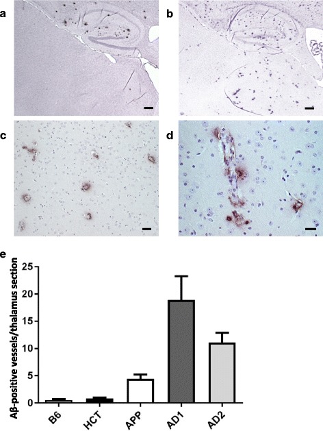
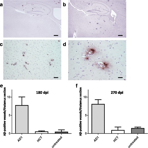
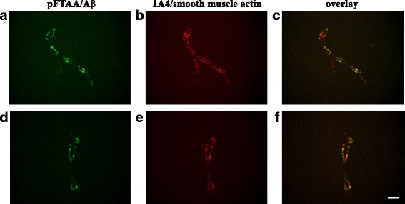
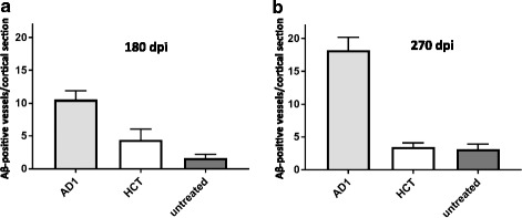
Seeding and spread of beta-amyloid (Aβ) pathologies have been considered to be based on prion-like mechanisms. However, limited transmissibility of Aβ seeding activity upon peripheral exposure would represent a key difference to prions, not only in terms of pathogenesis but also in terms of potential transmission of disease. We partially characterized the seeded Aβ amyloidosis after intracerebral injection of various brain homogenates in APP/PS1 mice. One particularly seed-laden homogenate was selected to investigate the development of Aβ pathologies after intravenous exposure. We report here that a single intravenous injection of an Alzheimer disease patient's-brain extract into APP/PS1 recipient mice led to cerebral amyloid angiopathy within 180 days post injection. Thus, vascular proteinopathies such as CAA are transmissible in mice via the intravenous route of peripheral exposure.


Conflict of interest statement

Competing interests

All authors declare that they have no competing interests.

Publisher’s Note

Springer Nature remains neutral with regard to jurisdictional claims in published maps and institutional affiliations.

Figures

Fig. 1
Fig. 1
Vascular amyloid deposition following intracerebral injection of brain extracts into APP/PS1 mice. Aβ deposits were detected 360 days post injection using the 4G8 monoclonal antibody. a Representative overview of the hippocampus and thalamus regions upon injection of the negative control homogenate HCT. b Hippocampus and thalamus regions after injection of AD1 homogenate. Scale bar 500 μm. c, d Examples of thalamic CAA after injection of the AD1 extract at higher magnifications. The majority of Aβ deposits in the thalamus is vascular. Scale bars 25 μm in (c) and 12.5 μm in (d). e Quantification of thalamic CAA 360 days after intracerebral injection of brain extracts. Indicated is the mean ± SEM. Mann-Whitney U test, group sizes n = 5 (HCT), n = 6 (B6), n = 6 (APP), n = 7 (AD1), and n = 7 (AD2). P = 0.003 for B6 versus AD1 and AD2; p = 0.0085 B6 versus APP, p = 0.38 B6 versus HCT, p = 0.1026 for AD1 versus AD2. Of note, the CAA in AD patients extracts AD1- and AD2-injected mice was significantly more pronounced than in mice receiving the APP/PS1 brain homogenate; p = 0.0264 and p = 0.0052 for AD1 and AD2 versus APP, respectively
Fig. 2
Fig. 2
CAA following intravenous injection of brain extracts into APP/PS1 mice. a Representative overview of the hippocampus and thalamus regions upon injection of the negative control homogenate HCT. b Hippocampus and thalamus regions after injection of AD patient homogenate AD1, 180 days post injection. Scale bar 500 μm. c Example of thalamic CAA after injection of the AD1 extract at higher magnification. The majority of Aβ deposits in the thalamus is vascular. Scale bar 25 μm. d Thalamic CAA with notable spread of Aβ deposition into the adjacent tissue after injection of the AD1 extract. Scale bar 12.5 μm. e Quantification of thalamic CAA 180 days after intravenous injection of brain extracts. Untreated APP/PS1 mice of the same age, which did not receive any injections, were included for additional comparison. Indicated is the mean ± SEM. Mann-Whitney U test, group sizes n = 6 (AD1), n = 6 (HCT) and n = 7 (untreated). P = 0.008 and p = 0.0062 for AD1 versus HCT and the untreated group of mice, respectively. f Quantification of thalamic CAA 270 days after intravenous injection of brain extracts. Indicated is the mean ± SEM. Kruskal-Wallis test, group sizes n = 3 (AD1), n = 3 (HCT) and n = 6 (untreated). P = 0.038, with p < 0.1 for AD1 versus HCT and the untreated group, respectively, and p > 0.1 for HCT versus untreated (Dunn’s multiple comparison test)
Fig. 3
Fig. 3
Double-stainings with amyloid-binding compound pFTAA and anti-smooth muscle actin antibody 1A4 to demonstrate the vascular Aβ deposition in thalami of intravenously AD1 injected mice. a, d staining with pFTAA; b, e staining of the same section with 1A4 antibody; c, f overlay of the pFTAA (green)/1A4 (red) stains. Scale bar 12.5 mm
Fig. 4
Fig. 4
CAA in cortices and cortically attached leptomeninges following intravenous injection of brain extracts into APP/PS1 mice. a CAA quantification 180 days after intravenous injection of brain extracts. Indicated is the mean ± SEM. Mann-Whitney U test, group sizes as in Fig. 2e. P = 0.027 and p = 0.016 for AD1 versus HCT and the untreated group of mice, respectively. b CAA quantification 270 days after intravenous injection of brain extracts. Kruskal-Wallis test, group sizes as in Fig. 2f. P = 0.0214, with p < 0.05 for AD1 versus the untreated group, and p > 0.1 for the other comparisons (Dunn’s multiple comparison test)

References

    1. Aslund A, Sigurdson CJ, Klingstedt T, Grathwohl S, Bolmont T, Dickstein DL et al (2009) Novel pentameric thiophene derivatives for in vitro and in vivo optical imaging of a plethora of protein aggregates in cerebral amyloidoses. ACS Chem Biol 4:673–684. - PMC - PubMed
    1. Brundin P, Melki R, Kopito R. Prion-like transmission of protein aggregates in neurodegenerative diseases. Nat Rev Mol Cell Biol. 2010;11:301–307. - PMC - PubMed
    1. Bu XL, Xiang Y, Jin WS, Wang J, Shen LL, Huang ZL et al (2017) Blood-derived amyloid-β protein induces Alzheimer’s disease pathologies. Mol Psychiatry. 10.1038/mp.2017.204. - PubMed
    1. Cohen ML, Kim C, Haldiman T, ElHag M, Mehndiratta P, Pichet T, et al. Rapidly progressive Alzheimer’s disease features distinct structures of amyloid-β. Brain. 2015;138:1009–1022. - PMC - PubMed
    1. Edgren G, Hjalgrim H, Rostgaard K, Erikstrup C, Sartipy U, Holzmann MJ, et al. Transmission of neurodegenerative disorders through blood transfusion: a cohort study. Ann Intern Med. 2016;165:316–324. - PubMed

Publication types

MeSH terms