. 2018 Mar 5;37(1):45.
doi: 10.1186/s13046-018-0720-8.

Downregulation of castor zinc finger 1 predicts poor prognosis and facilitates hepatocellular carcinoma progression via MAPK/ERK signaling

Affiliations

Downregulation of castor zinc finger 1 predicts poor prognosis and facilitates hepatocellular carcinoma progression via MAPK/ERK signaling

Ji-Long Wang et al. J Exp Clin Cancer Res. .

Abstract

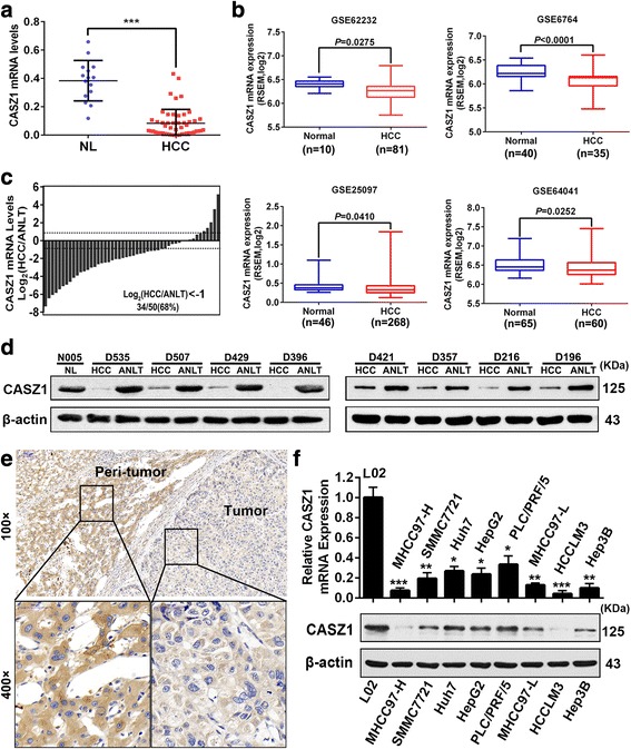
Background: Castor zinc finger 1 (CASZ1) plays critical roles in various biological processes and pathologic conditions, including cancer. However, the prognostic importance and biologic functions of CASZ1 in hepatocellular carcinoma (HCC) are still unclear.

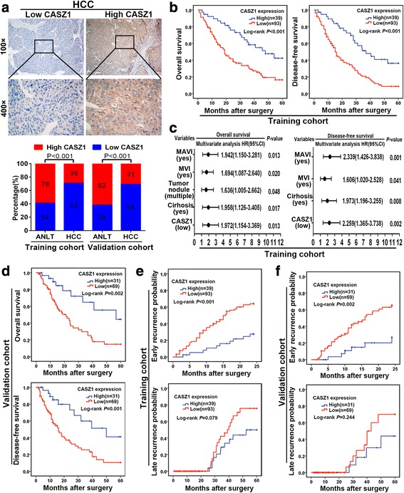
Methods: qRT-PCR, western blot and immunohistochemistry analyses were used to determine CASZ1 expression in HCC samples and cell lines. The clinical significance of CASZ1 was assessed in two independent study cohorts containing 232 patients with HCC. A series of in vitro and in vivo experiments were performed to explore the role and molecular mechanism of CASZ1 in HCC progression.

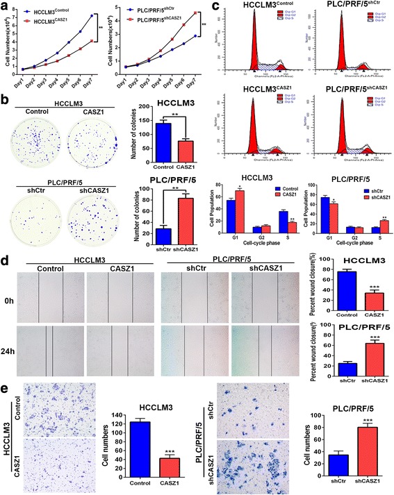
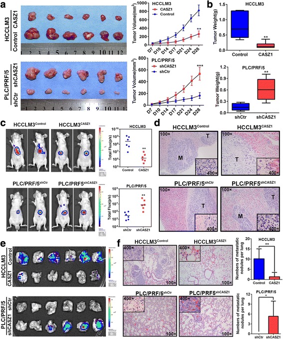
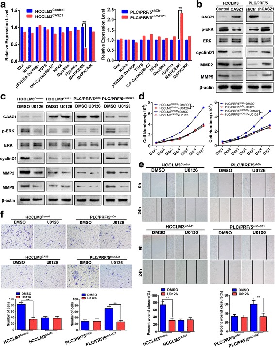
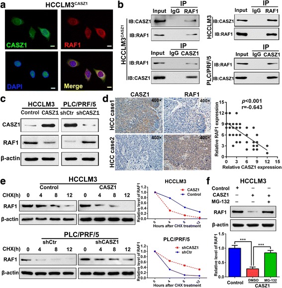
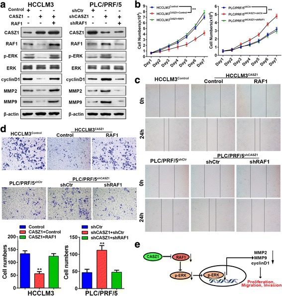
Results: Here we report that CASZ1 expression was downregulated in HCC tissues and cell lines. Low CASZ1 expression was closely correlated with aggressive clinicopathological features, poor clinical outcomes and early recurrence of HCC patients. Moreover, overexpression of CASZ1 in HCCLM3 cells significantly inhibited cell proliferation, migration, invasion in vitro and tumor growth and metastasis in vivo, whereas silencing CASZ1 significantly enhanced the above abilities of PLC/PRF/5 cells. Further mechanism study indicated that these phenotypic changes were mediated by MAPK/ERK signaling pathway and involved altered expression of MMP2, MMP9 and cyclinD1. Finally, we proved that CASZ1 exerted its tumor-suppressive effect by directly interacting with RAF1 and reducing the protein stability of RAF1.

Conclusions: Our study for the first time demonstrated that CASZ1 is a tumor suppressor in HCC, which may serve as a novel prognostic predictor and therapeutic target for HCC patients.

Keywords: Castor zinc finger 1; Hepatocellular carcinoma; MAPK/ERK; Progression; RAF1.


Conflict of interest statement

Ethics approval and consent to participate

The studies were approved by the Ethics Committee of Xiangya Hospital of Central South University and Affiliated Cancer Hospital of Xiangya School of Medicine. Written informed consent was obtained from all patients. Animal experiments were approved by the Institutional Animal Care and Use Committee of Central South University.

Consent for publication

Not applicable

Competing interests

The authors declare that they have no competing interests.

Publisher’s Note

Springer Nature remains neutral with regard to jurisdictional claims in published maps and institutional affiliations.

Figures

Fig. 1
Fig. 1
CASZ1 is downregulated in human HCC tissues and cell lines a. CASZ1 mRNA expression in 15 normal liver tissues (NLs) and 50 HCC tissues was analyzed by qRT-PCR. Data are shown as mean ± SD. ***P < 0.001. b CASZ1 expression was lower in HCC tissues than NLs according to the analysis of data from GEO (GSE62232, GSE6764, GSE25097, GSE64041, all P < 0.05). c Waterfall plot showing the downregulation of CASZ1 in 34 of 50 (68%) HCC samples compared to their matched adjacent non-tumor liver tissues (ANLTs). d The CASZ1 protein levels in HCCs, ANLTs and NLs were analyzed by Western blot. β-actin was used as a loading control. e Representative immunohistochemical images demonstrated CASZ1 protein was lowly expressed in HCC tumor tissues compared with their peri-tumor tissues. Magnification, × 100, × 400. f Expression of CASZ1 in HCC cell lines and L02, the normal liver cell line, were measured by qRT-PCR and western blot, respectively. *P < 0.05; **P < 0.01; ***P < 0.001 compared with L02
Fig. 2
Fig. 2
Low expression of CASZ1 is associated with aggressive clinicopathological characteristics and poor prognosis a. Representative images of low CASZ1 expression cases and high CASZ1 expression cases were shown (upper panel). Magnification, × 100, × 400. The percentages of low or high CASZ1 in paired HCC samples from the training and validation cohorts were compared (lower panel). b Kaplan-Meier analysis of OS and DFS based on CASZ1 expression in the training cohort. c Forest plots showing HR of OS and DFS for HCC patients in the indicated clinical subgroups of training cohort. d Kaplan-Meier analysis of OS and DFS based on CASZ1 expression in the validation cohort. e Kaplan-Meier analysis of early recurrence and late recurrence based on CASZ1 expression in the training cohort. f Kaplan-Meier analysis of early recurrence and late recurrence based on CASZ1 expression in the validation cohort
Fig. 3
Fig. 3
CASZ1 inhibits HCC cell proliferation, migration and invasion in vitro a. The effects of CASZ1 on HCC cell proliferation was measured by MTT assay at different time points. b Representative imgages and quantification of cell clones in HCCLM3CASZ1, PLC/PRF/5shCASZ1 and their control groups, as determined by colony formation assay. c The effect of CASZ1 on HCC cell cycle progression was analyzed by flow cytometry. d Wound healing assay was performed to detect the migratory capacity of HCCLM3CASZ1, PLC/PRF/5shCASZ1 and their control cells. e Transwell invasion assay was performed to evaluate the invasive potential of CASZ1-interfered HCC cells. Each bar represents the mean ± SD of three independent experiments. *P < 0.05; **P < 0.01; ***P < 0.001
Fig. 4
Fig. 4
CASZ1 represses HCC growth and metastasis in vivo a. Effects of CASZ1 on HCC growth in subcutaneous xenograft model. The size of tumors was measured at indicated time points, and is shown as mean ± SD. **P < 0.01; ***P < 0.001. b Tumors were excised and weighted after mice were sacrificed. **P < 0.01. c Representative bioluminescent images of the orthotopic HCC model in HCCLM3Control, HCCLM3CASZ1, PLC/PRF/5shCtr and PLC/PRF/5shCASZ1 groups (n = 6 for each group, left panel). The colored region represents the fluorescence signal of HCC cells in nude mice. Luciferase activity was quantified for experimental and control groups (right panel). **P < 0.01. d Representative H&E-stained sections of orthotopic primary liver tumors formed by HCCLM3CASZ1, PLC/PRF/5shCASZ1 and their control cells (original magnified × 100; inserted figures magnified × 400). e The presence of lung metastasis was examined in ex vivo bioluminescence imaging. f Representative H&E staining of pulmonary metastatic nodules in experimental and control groups (left panel: original magnified × 100; inserted figures magnified × 400). The number of lung metastatic nodules in each group is presented as mean ± SD (right panel). *P < 0.05; **P < 0.01
Fig. 5
Fig. 5
CASZ1 inhibits HCC growth and metastasis via inactivating the MAPK/ERK signaling a. 10-Pathway Reporter Array was performed to detect the signaling changes in CASZ1-interfered HCC cells. **P < 0.01. b The expression of critical members and downstream effectors of MAPK/ERK pathway was examined by western blot in HCCLM3CASZ1, PLC/PRF/5shCASZ1 and their control cells. c The levels of p-ERK, MMP2, MMP9 and cyclinD1 were determined by western blot in HCCLM3CASZ1, PLC/PRF/5shCASZ1 and their control cells treated with U0126 (10 μM) or DMSO control. d-f. HCCLM3CASZ1, PLC/PRF/5shCASZ1 and their control cells were treated with U0126 (10 μM) or DMSO control and then subjected to MTT (d), wound healing (e) and transwell invasion (f) assays
Fig. 6
Fig. 6
CASZ1 decreases RAF1 expression by reducing the protein stability of RAF1 a. HCCLM3 cells transfected with CASZ1 were fixed for the double immunofluorescence staining, and colocalization of CASZ1 with RAF1 were visualized as yellow fluorescence in merge panel. Scale bar, 25 μm. b Co-immunoprecipitation assay was performed to analyze the direct binding between CASZ1 and RAF1 in HCCLM3CASZ1, HCCLM3 and PLC/PRF/5 cells. c The expression of RAF1 was determined in CASZ1-interfered HCC cells by western blot. d Correlation between CASZ1 and RAF1 was measured in 50 HCC samples. Representative IHC staining of CASZ1 and RAF1 (left panel, magnification, × 400) in serial sections are shown, indicating a negative correlation between the protein level of CASZ1 and RAF1 in the clinical samples (r = − 0.643, P < 0.001, right panel). e The half-life of RAF1 protein in HCC cells was analyzed following treatment with cycloheximide (CHX, 25 μg/ml) for the indicated time points. The half-life of RAF1 protein was decreased in HCCLM3 cells with CASZ1 overexpression, but increased in PLC/PRF/5 cells with CASZ1 knockdown. f MG-132 (20 μM) was used to inhibit the proteasomal degradation in HCCLM3 cells. MG-132 treatment reversed the downregulation of RAF1 protein induced by CASZ1 overexpression. *** P < 0.001
Fig. 7
Fig. 7
RAF1 is critical for CASZ1-mediated inhibition of MAPK/ERK signaling and HCC progression a. Western blot analysis of CASZ1, RAF1, p-ERK, MMP2, MMP9 and cyclinD1 expression in CASZ1-interfered HCC cells with RAF1 knockdown or ectopic expression. b-d. MTT (b), wound healing (c) and transwell invasion (d) assays were performed to test the influence of RAF1 on the proliferation, migration and invasion abilities of CASZ1-interfered HCC cells with RAF1 knockdown or ectopic expression. e Schematic depiction of the mechanism underlying CASZ1-mediated inhibition of HCC cell proliferation, migration and invasion based on our study

Similar articles

Cited by

References

    1. Chen W, Zheng R, Baade PD, Zhang S, Zeng H, Bray F, Jemal A, Yu XQ, He J. Cancer statistics in China, 2015. CA Cancer J Clin. 2016;66:115–132. doi: 10.3322/caac.21338. - DOI - PubMed
    1. Torre LA, Bray F, Siegel RL, Ferlay J, Lortet-Tieulent J, Jemal A. Global cancer statistics, 2012. CA Cancer J Clin. 2015;65:87–108. doi: 10.3322/caac.21262. - DOI - PubMed
    1. Forner A, Llovet JM, Bruix J. Hepatocellular carcinoma. Lancet. 2012;379:1245–1255. doi: 10.1016/S0140-6736(11)61347-0. - DOI - PubMed
    1. El-Serag HB. Hepatocellular carcinoma. N Engl J Med. 2011;365:1118–1127. doi: 10.1056/NEJMra1001683. - DOI - PubMed
    1. Xia L, Huang W, Tian D, Zhu H, Qi X, Chen Z, Zhang Y, Hu H, Fan D, Nie Y, Wu K. Overexpression of forkhead box C1 promotes tumor metastasis and indicates poor prognosis in hepatocellular carcinoma. Hepatology. 2013;57:610–624. doi: 10.1002/hep.26029. - DOI - PubMed

MeSH terms