

TRPM2 Channel Aggravates CNS Inflammation and Cognitive Impairment via Activation of Microglia in Chronic Cerebral Hypoperfusion
- PMID: 29507145
- PMCID: PMC6596050
- DOI: 10.1523/JNEUROSCI.2451-17.2018
TRPM2 Channel Aggravates CNS Inflammation and Cognitive Impairment via Activation of Microglia in Chronic Cerebral Hypoperfusion
Abstract
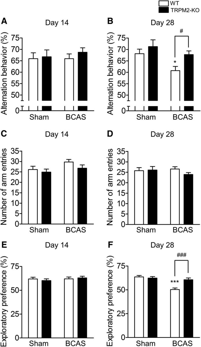
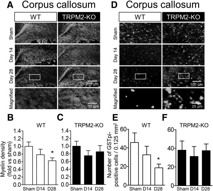
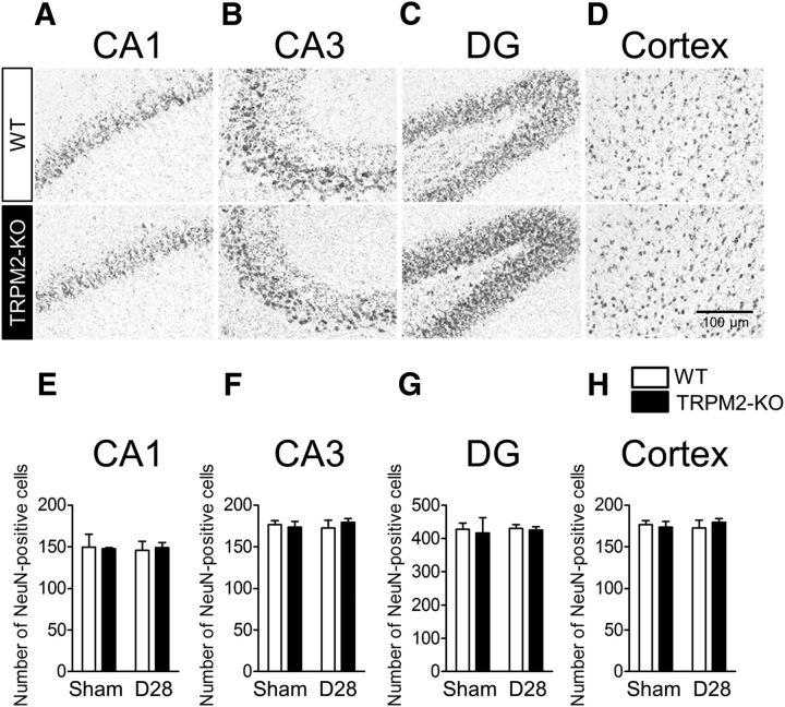
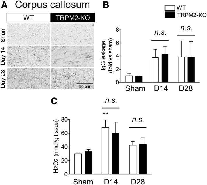
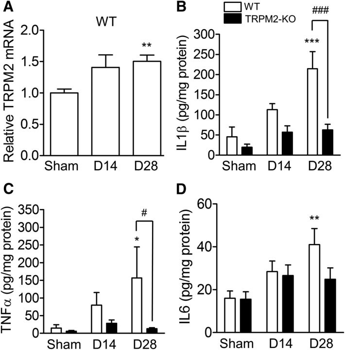
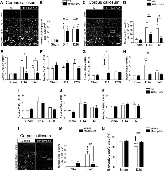
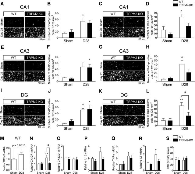
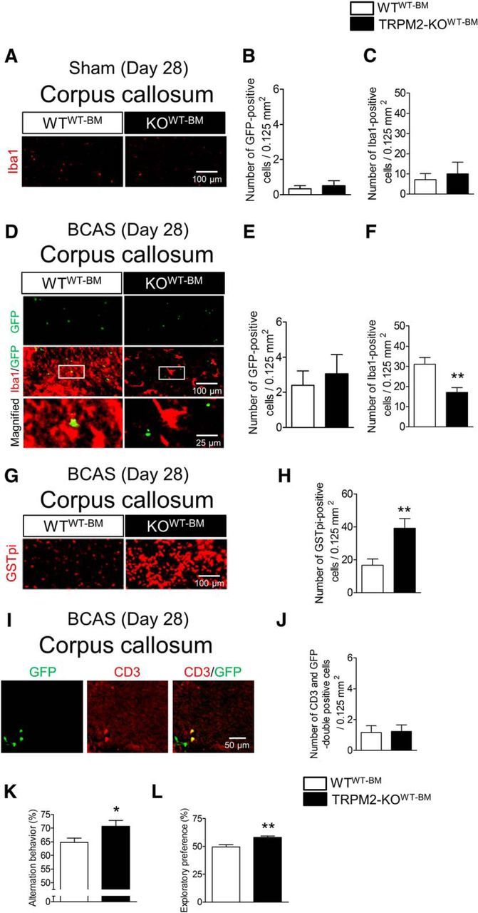
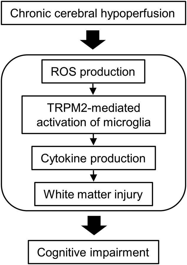
Chronic cerebral hypoperfusion is a characteristic seen in widespread CNS diseases, including neurodegenerative and mental disorders, and is commonly accompanied by cognitive impairment. Recently, several studies demonstrated that chronic cerebral hypoperfusion can induce the excessive inflammatory responses that precede neuronal dysfunction; however, the precise mechanism of cognitive impairment due to chronic cerebral hypoperfusion remains unknown. Transient receptor potential melastatin 2 (TRPM2) is a Ca2+-permeable channel that is abundantly expressed in immune cells and is involved in aggravation of inflammatory responses. Therefore, we investigated the pathophysiological role of TRPM2 in a mouse chronic cerebral hypoperfusion model with bilateral common carotid artery stenosis (BCAS). When male mice were subjected to BCAS, cognitive dysfunction and white matter injury at day 28 were significantly improved in TRPM2 knock-out (TRPM2-KO) mice compared with wild-type (WT) mice, whereas hippocampal damage was not observed. There were no differences in blood-brain barrier breakdown and H2O2 production between the two genotypes at 14 and 28 d after BCAS. Cytokine production was significantly suppressed in BCAS-operated TRPM2-KO mice compared with WT mice at day 28. In addition, the number of Iba1-positive cells gradually decreased from day 14. Moreover, daily treatment with minocycline significantly improved cognitive perturbation. Surgical techniques using bone marrow chimeric mice revealed that activated Iba1-positive cells in white matter could be brain-resident microglia, not peripheral macrophages. Together, these findings suggest that microglia contribute to the aggravation of cognitive impairment by chronic cerebral hypoperfusion, and that TRPM2 may be a potential target for chronic cerebral hypoperfusion-related disorders.SIGNIFICANCE STATEMENT Chronic cerebral hypoperfusion is manifested in a wide variety of CNS diseases, including neurodegenerative and mental disorders that are accompanied by cognitive impairment; however, the underlying mechanisms require clarification. Here, we used a chronic cerebral hypoperfusion mouse model to investigate whether TRPM2, a Ca2+-permeable cation channel highly expressed in immune cells, plays a destructive role in the development of chronic cerebral hypoperfusion-induced cognitive impairment, and propose a new hypothesis in which TRPM2-mediated activation of microglia, not macrophages, specifically contributes to the pathology through the aggravation of inflammatory responses. These findings shed light on the understanding of the mechanisms of chronic cerebral hypoperfusion-related inflammation, and are expected to provide a novel therapeutic molecule for cognitive impairment in CNS diseases.
Keywords: TRPM2; cerebral hypoperfusion; cognitive impairment; cytokine; microglia; white matter injury.
Copyright © 2018 the authors 0270-6474/18/383521-14$15.00/0.
Conflict of interest statement
The authors declare no competing financial interests.
Figures
Similar articles
-
Pathophysiological Role of TRPM2 in Age-Related Cognitive Impairment in Mice.Neuroscience. 2019 Jun 1;408:204-213. doi: 10.1016/j.neuroscience.2019.04.012. Epub 2019 Apr 15. Neuroscience. 2019. PMID: 30999030
-
Depletion of microglia ameliorates white matter injury and cognitive impairment in a mouse chronic cerebral hypoperfusion model.Biochem Biophys Res Commun. 2019 Jul 5;514(4):1040-1044. doi: 10.1016/j.bbrc.2019.05.055. Epub 2019 May 13. Biochem Biophys Res Commun. 2019. PMID: 31097227
-
Deficiency of Nrf2 exacerbates white matter damage and microglia/macrophage levels in a mouse model of vascular cognitive impairment.J Neuroinflammation. 2020 Dec 1;17(1):367. doi: 10.1186/s12974-020-02038-2. J Neuroinflammation. 2020. PMID: 33261626 Free PMC article.
-
[Roles of transient receptor potential melastatin 2 expressed on immune cells in neuropathic pain].Yakugaku Zasshi. 2014;134(3):379-86. doi: 10.1248/yakushi.13-00236-2. Yakugaku Zasshi. 2014. PMID: 24584019 Review. Japanese.
-
A brief overview of a mouse model of cerebral hypoperfusion by bilateral carotid artery stenosis.J Cereb Blood Flow Metab. 2023 Nov;43(2_suppl):18-36. doi: 10.1177/0271678X231154597. Epub 2023 Mar 8. J Cereb Blood Flow Metab. 2023. PMID: 36883344 Free PMC article. Review.
Cited by
-
TRPM2 Channel in Microglia as a New Player in Neuroinflammation Associated With a Spectrum of Central Nervous System Pathologies.Front Pharmacol. 2019 Mar 12;10:239. doi: 10.3389/fphar.2019.00239. eCollection 2019. Front Pharmacol. 2019. PMID: 30914955 Free PMC article. Review.
-
Aberrant oligodendroglial LDL receptor orchestrates demyelination in chronic cerebral ischemia.J Clin Invest. 2021 Jan 4;131(1):e128114. doi: 10.1172/JCI128114. J Clin Invest. 2021. PMID: 33141760 Free PMC article.
-
Targeting NLRP3 signaling with a novel sulfonylurea compound for the treatment of vascular cognitive impairment and dementia.Res Sq [Preprint]. 2024 Dec 20:rs.3.rs-5611378. doi: 10.21203/rs.3.rs-5611378/v1. Res Sq. 2024. Update in: Fluids Barriers CNS. 2025 Jun 3;22(1):55. doi: 10.1186/s12987-025-00665-6. PMID: 39764140 Free PMC article. Updated. Preprint.
-
Integrative Multiomics Profiling of Mouse Hippocampus Reveals Transcriptional Upregulation of Interferon-Stimulated Genes Through PU.1 Regulator in Microglial Activation Induced by Chronic Cerebral Hypoperfusion.MedComm (2020). 2025 Apr 15;6(5):e70157. doi: 10.1002/mco2.70157. eCollection 2025 May. MedComm (2020). 2025. PMID: 40242160 Free PMC article.
-
A Systemic Review of the Integral Role of TRPM2 in Ischemic Stroke: From Upstream Risk Factors to Ultimate Neuronal Death.Cells. 2022 Jan 31;11(3):491. doi: 10.3390/cells11030491. Cells. 2022. PMID: 35159300 Free PMC article.
References
-
- Aquila R, Citrome L (2015) Cognitive impairment in schizophrenia: the great unmet need. CNS Spectr 20:35–39; quiz 40. - PubMed
Publication types
MeSH terms
Substances
Grants and funding
LinkOut - more resources
Full Text Sources
Other Literature Sources
Molecular Biology Databases
Research Materials
Miscellaneous