

B7-H1 maintains the polyclonal T cell response by protecting dendritic cells from cytotoxic T lymphocyte destruction
- PMID: 29507197
- PMCID: PMC5866601
- DOI: 10.1073/pnas.1722043115
B7-H1 maintains the polyclonal T cell response by protecting dendritic cells from cytotoxic T lymphocyte destruction
Erratum in
-
Correction for Chen et al., B7-H1 maintains the polyclonal T cell response by protecting dendritic cells from cytotoxic T lymphocyte destruction.Proc Natl Acad Sci U S A. 2018 Apr 17;115(16):E3857. doi: 10.1073/pnas.1804362115. Epub 2018 Apr 2. Proc Natl Acad Sci U S A. 2018. PMID: 29610321 Free PMC article. No abstract available.
Abstract
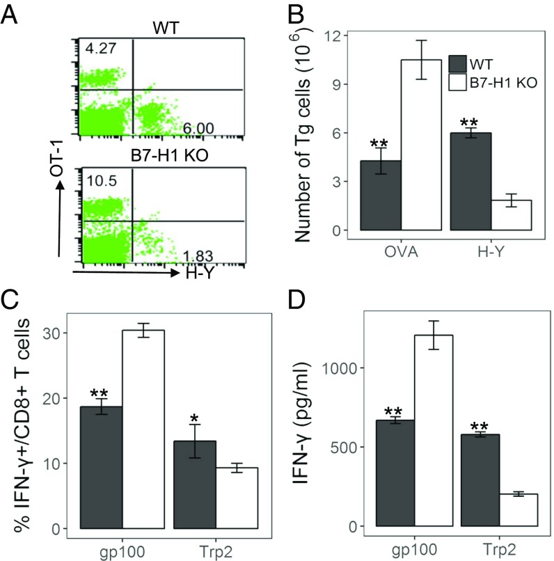
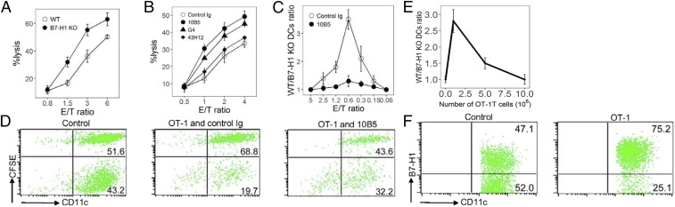
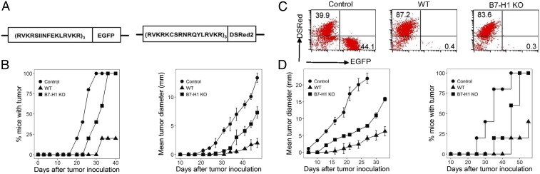
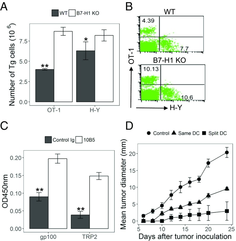
Induced B7-H1 expression in the tumor microenvironment initiates adaptive resistance, which impairs immune functions and leads to tumor escape from immune destruction. Antibody blockade of the B7-H1/PD-1 interaction overcomes adaptive resistance, leading to regression of advanced human cancers and survival benefits in a significant fraction of patients. In addition to cancer cells, B7-H1 is expressed on dendritic cells (DCs), but its role in DC functions is less understood. DCs can present multiple antigens (Ags) to stimulate dominant or subdominant T cell responses. Here, we show that immunization with multiple tumor Ag-loaded DCs, in the absence of B7-H1, vastly enhances cytotoxic T lymphocyte (CTL) responses to dominant Ag. In sharp contrast, CTL responses to subdominant Ag were paradoxically suppressed, facilitating outgrowth of tumor variants carrying only subdominant Ag. Suppressed CTL responses to subdominant Ag are largely due to the loss of B7-H1-mediated protection of DCs from the lysis of CTL against dominant Ag. Therefore, B7-H1 expression on DCs may help maintain the diversity of CTL responses to multiple tumor Ags. Interestingly, a split immunization approach, which presents dominant and subdominant Ags with different DCs, promoted CTL responses to all Ags and prevented tumor escape in murine tumor models. These findings have implications for the design of future combination cancer immunotherapies.
Keywords: B7-H1; PD-1; cytolytic T cells; dominant antigen; subdominant antigen.
Conflict of interest statement
Conflict of interest statement: Lieping Chen is a consultant/advisory board member/receives consulting fees from MedImmune, Pfizer, NextCure, Vcanbio, and GenomiCare and currently has sponsored research grants from Boehringer Ingelheim, Pfizer, and NextCure.
Figures
Similar articles
-
Expression of programmed death 1 ligands by murine T cells and APC.J Immunol. 2002 Nov 15;169(10):5538-45. doi: 10.4049/jimmunol.169.10.5538. J Immunol. 2002. PMID: 12421930
-
Blockade of B7-H1 enhances dendritic cell-mediated T cell response and antiviral immunity in HBV transgenic mice.Vaccine. 2012 Jan 17;30(4):758-66. doi: 10.1016/j.vaccine.2011.11.076. Epub 2011 Nov 29. Vaccine. 2012. PMID: 22133510
-
Blockade of B7-H1 and PD-1 by monoclonal antibodies potentiates cancer therapeutic immunity.Cancer Res. 2005 Feb 1;65(3):1089-96. Cancer Res. 2005. PMID: 15705911
-
Tumor-infiltrating programmed death receptor-1+ dendritic cells mediate immune suppression in ovarian cancer.J Immunol. 2011 Jun 15;186(12):6905-13. doi: 10.4049/jimmunol.1100274. Epub 2011 May 6. J Immunol. 2011. PMID: 21551365 Free PMC article.
-
T cell costimulation by B7/BB1 induces CD8 T cell-dependent tumor rejection: an important role of B7/BB1 in the induction, recruitment, and effector function of antitumor T cells.J Exp Med. 1994 Apr 1;179(4):1205-14. doi: 10.1084/jem.179.4.1205. J Exp Med. 1994. PMID: 7511683 Free PMC article.
Cited by
-
CD80-Fc fusion protein as a potential cancer immunotherapy strategy.Antib Ther. 2023 Nov 30;7(1):28-36. doi: 10.1093/abt/tbad029. eCollection 2024 Jan. Antib Ther. 2023. PMID: 38235375 Free PMC article. Review.
-
Neoantigen architectures define immunogenicity and drive immune evasion of tumors with heterogenous neoantigen expression.J Immunother Cancer. 2024 Nov 9;12(11):e010249. doi: 10.1136/jitc-2024-010249. J Immunother Cancer. 2024. PMID: 39521615 Free PMC article.
-
PD-1/PD-L1 co-inhibition shapes anticancer T cell immunodominance: facing the consequences of an immunological ménage à trois.Cancer Immunol Immunother. 2018 Nov;67(11):1669-1672. doi: 10.1007/s00262-018-2231-z. Epub 2018 Aug 21. Cancer Immunol Immunother. 2018. PMID: 30132082 Free PMC article.
-
Synergistic anti-tumor effect of anti-PD-L1 antibody cationic microbubbles for delivery of the miR-34a gene combined with ultrasound on cervical carcinoma.Am J Transl Res. 2021 Mar 15;13(3):988-1005. eCollection 2021. Am J Transl Res. 2021. PMID: 33841635 Free PMC article.
-
PD-L1 on dendritic cells attenuates T cell activation and regulates response to immune checkpoint blockade.Nat Commun. 2020 Sep 24;11(1):4835. doi: 10.1038/s41467-020-18570-x. Nat Commun. 2020. PMID: 32973173 Free PMC article.
References
-
- Yewdell JW, Bennink JR. Immunodominance in major histocompatibility complex class I-restricted T lymphocyte responses. Annu Rev Immunol. 1999;17:51–88. - PubMed
-
- Yewdell JW. Confronting complexity: Real-world immunodominance in antiviral CD8+ T cell responses. Immunity. 2006;25:533–543. - PubMed
-
- Halstead ES, Mueller YM, Altman JD, Katsikis PD. In vivo stimulation of CD137 broadens primary antiviral CD8+ T cell responses. Nat Immunol. 2002;3:536–541. - PubMed
-
- Cheuk E, et al. Human MHC class I transgenic mice deficient for H2 class I expression facilitate identification and characterization of new HLA class I-restricted viral T cell epitopes. J Immunol. 2002;169:5571–5580. - PubMed
Publication types
MeSH terms
Substances
Grants and funding
LinkOut - more resources
Full Text Sources
Other Literature Sources
Molecular Biology Databases
Research Materials
