. 2018 Mar 20;115(12):3036-3041.
doi: 10.1073/pnas.1800756115. Epub 2018 Mar 5.

Mechanism of the G-protein mimetic nanobody binding to a muscarinic G-protein-coupled receptor

Affiliations

Mechanism of the G-protein mimetic nanobody binding to a muscarinic G-protein-coupled receptor

Yinglong Miao et al. Proc Natl Acad Sci U S A. .

Abstract

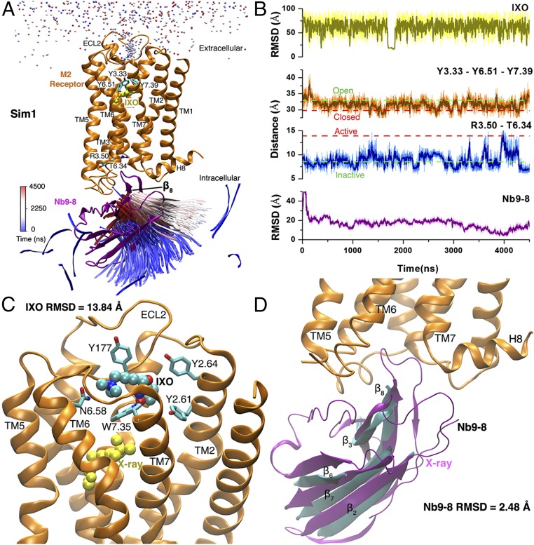
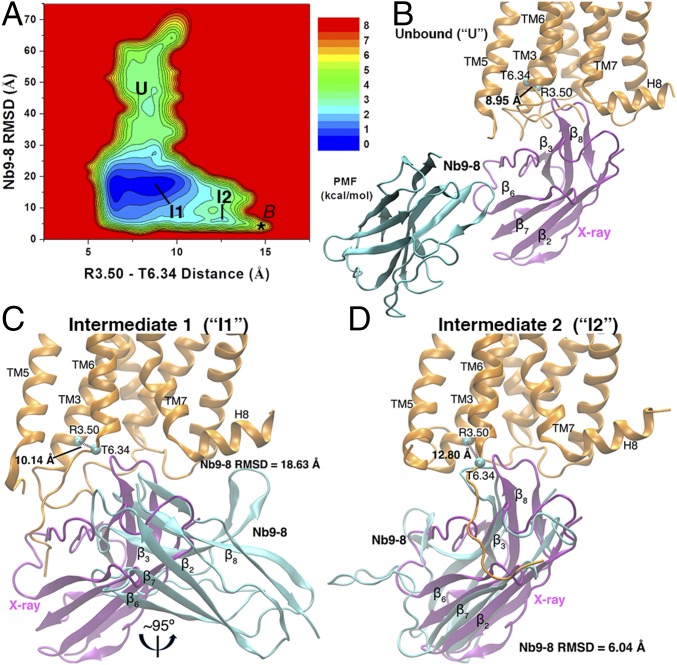
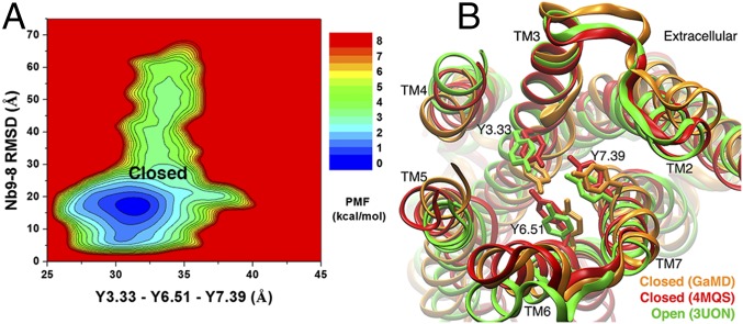
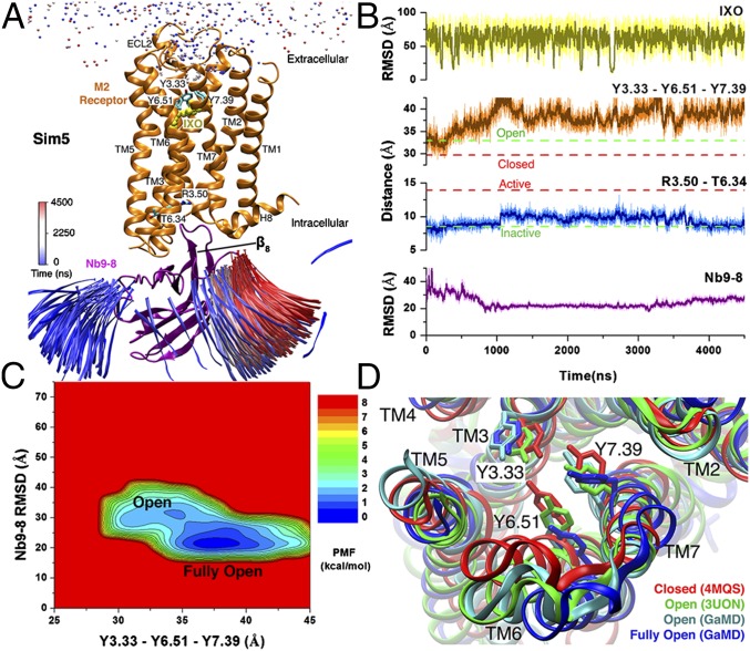
Protein-protein binding is key in cellular signaling processes. Molecular dynamics (MD) simulations of protein-protein binding, however, are challenging due to limited timescales. In particular, binding of the medically important G-protein-coupled receptors (GPCRs) with intracellular signaling proteins has not been simulated with MD to date. Here, we report a successful simulation of the binding of a G-protein mimetic nanobody to the M2 muscarinic GPCR using the robust Gaussian accelerated MD (GaMD) method. Through long-timescale GaMD simulations over 4,500 ns, the nanobody was observed to bind the receptor intracellular G-protein-coupling site, with a minimum rmsd of 2.48 Å in the nanobody core domain compared with the X-ray structure. Binding of the nanobody allosterically closed the orthosteric ligand-binding pocket, being consistent with the recent experimental finding. In the absence of nanobody binding, the receptor orthosteric pocket sampled open and fully open conformations. The GaMD simulations revealed two low-energy intermediate states during nanobody binding to the M2 receptor. The flexible receptor intracellular loops contribute remarkable electrostatic, polar, and hydrophobic residue interactions in recognition and binding of the nanobody. These simulations provided important insights into the mechanism of GPCR-nanobody binding and demonstrated the applicability of GaMD in modeling dynamic protein-protein interactions.

Keywords: GPCR signaling; biomolecular recognition; enhanced sampling; pathways; protein binding.


Conflict of interest statement

The authors declare no conflict of interest.

Figures

Fig. 1.
Fig. 1.
Binding of agonist IXO and G-protein mimetic nanobody Nb9-8 to the M2 receptor was observed in one 4,500-ns GaMD simulation. (A) Trajectories of nitrogen in the trimethylamine group of IXO (beads) and the β8 strand of Nb9-8 (ribbons) colored by simulation time in a blue (0 ns)–white (2,250 ns)–red (4,500 ns) scale. The β8 strand of Nb9-8 moves into the X-ray conformation near the simulation end. X-ray conformations of the M2 receptor and Nb9-8 (PDB ID code 4MQS) are shown in orange and purple ribbons, respectively, and yellow spheres for IXO. Residues Tyr1043.33, Tyr4036.51, Tyr4267.39, Arg1213.50, and Thr3866.34 are represented by sticks. (B) The rmsds of the IXO and Nb9-8 relative to the X-ray structure, Tyr1043.33−Tyr4036.51−Tyr4267.39 triangle perimeter and Arg1213.50−Thr3866.34 distance calculated from the simulation. Dashed lines indicate X-ray structural values of the M2 receptor (3UON, green; 4MQS, red). (C) Binding pose of IXO (spheres) in the receptor extracellular vestibule with 13.84-Å rmsd relative to the X-ray conformation (yellow spheres). Residues found within 5 Å of IXO are highlighted in sticks. (D) Binding pose of Nb9-8 (cyan) in the receptor intracellular pocket with 2.48-Å rmsd relative to the X-ray conformation (purple). The β2, β3, β6, β7, and β8 strands that represent the nanobody core domain were selected for calculating rmsd of Nb9-8 and heavy atoms for IXO.
Fig. 2.
Fig. 2.
(A) The 2D PMF calculated with the Arg1213.50−Thr3866.34 distance and rmsd of the Nb9-8 relative to the 4MQS X-ray conformation. Three low-energy conformational states are labeled as the Unbound (U), Intermediate 1 (I1), and Intermediate 2 (I2). The bound (B) state obtained from previous GaMD simulation of the 4MQS X-ray structure (29) is labeled a star. (BD) Structural conformations of the M2 receptor and Nb9-8 in the U (B), I1 (C), and I2 (D) states, for which energy minima of the (Arg1213.50−Thr3866.34 distance, Nb9-8 rmsd) 2D PMF were found at (8.95 Å, 49.44 Å), (10.14 Å, 18.63 Å), and (12.80 Å, 6.04 Å), respectively. The reference X-ray conformations of the M2 receptor and nanobody are shown as orange and purple ribbons, respectively. The evolving nanobody is represented by cyan ribbons.
Fig. 3.
Fig. 3.
Residue interactions between the M2 receptor and the G-protein mimetic nanobody Nb9-8 in the low-energy conformational states. (A and B) The I1 conformation looking at the TM5–TM6–TM7 (A) and TM1–TM2–TM4 (B) helices. (C and D) The I2 conformation looking at the TM5–TM6–TM7 (C) and TM1–TM2–TM4 (D) helices. (E and F) The B conformation looking at the TM5–TM6–TM7 (E) and TM1–TM2–TM4 (F) helices. The M2 receptor is shown in orange ribbons, and residues found within 2.5 Å of the nanobody are represented by sticks. The nanobody is represented by surface and colored by the atomic charges in a blue (+0.5e)–white (0)–red (−0.5e) scale. Key residues in the nanobody that interact with the M2 receptor are labeled in purple.
Fig. 4.
Fig. 4.
(A) The 2D PMF calculated by using the Sim1 GaMD trajectory regarding the Tyr1043.33−Tyr4036.51−Tyr4267.39 triangle perimeter and rmsd of Nb9-8 relative to the 4MQS X-ray conformation. The low-energy conformational state is labeled as the closed. (B) Extracellular view of the GaMD simulation-derived closed conformational state (orange) with the closed (red) and open (green) X-ray conformations (PDB ID codes 4MQS and 3UON). The M2 receptor is represented by ribbons, and residues Tyr1043.33, Tyr4036.51, and Tyr4267.39 are highlighted in sticks.
Fig. 5.
Fig. 5.
One 4,500-ns GaMD simulation of the M2 receptor (Sim5 in Table 1), during which IXO and Nb9-8 bind to the receptor with the smallest rmsds of 8.03 and 15.71 Å, respectively, relative to the 4MQS X-ray conformation. (A) Trajectories of nitrogen in the trimethylamine group of IXO (beads) and the β8 strand of Nb9-8 (ribbons) colored by the simulation time in a blue (0 ns)–white (2,250 ns)–red (4,500 ns) scale. The structural representations are similar to Fig. 1A. (B) The rmsds of the IXO and Nb9-8 relative to the 4MQS X-ray conformations, Tyr1043.33−Tyr4036.51−Tyr4267.39 triangle perimeter, and Arg1213.50−Thr3866.34 distance calculated from the simulation. Dashed lines indicate X-ray structural values of the M2 receptor (3UON, green; 4MQS, red). (C) The 2D PMF calculated with the Tyr1043.33−Tyr4036.51−Tyr4267.39 triangle perimeter and rmsd of the Nb9-8 relative to the 4MQS X-ray conformation. Two low-energy conformational states are labeled as open and fully open. (D) Extracellular view of the GaMD simulation-derived open (cyan) and fully open (blue) conformational states compared with the closed (red) and open (green) X-ray conformational states (PDB ID codes 4MQS and 3UON). The M2 receptor is represented by ribbons, and residues Tyr1043.33, Tyr4036.51, and Tyr4267.39 are highlighted in sticks.

References

    1. Hauser AS, Attwood MM, Rask-Andersen M, Schiöth HB, Gloriam DE. Trends in GPCR drug discovery: New agents, targets and indications. Nat Rev Drug Discov. 2017;16:829–842. - PMC - PubMed
    1. Kruse AC, et al. Muscarinic acetylcholine receptors: Novel opportunities for drug development. Nat Rev Drug Discov. 2014;13:549–560. - PMC - PubMed
    1. Spalding TA, Burstein ES. Constitutive activity of muscarinic acetylcholine receptors. J Recept Signal Transduct Res. 2006;26:61–85. - PubMed
    1. Haga K, et al. Structure of the human M2 muscarinic acetylcholine receptor bound to an antagonist. Nature. 2012;482:547–551. - PMC - PubMed
    1. Kruse AC, et al. Activation and allosteric modulation of a muscarinic acetylcholine receptor. Nature. 2013;504:101–106. - PMC - PubMed

Publication types

MeSH terms

Substances