. 2018 Mar 20;115(12):E2716-E2724.
doi: 10.1073/pnas.1719110115. Epub 2018 Mar 5.

N-terminal arginylation generates a bimodal degron that modulates autophagic proteolysis

Affiliations

N-terminal arginylation generates a bimodal degron that modulates autophagic proteolysis

Young Dong Yoo et al. Proc Natl Acad Sci U S A. .

Abstract

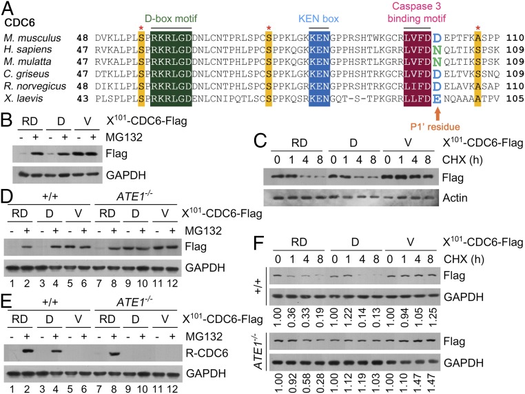
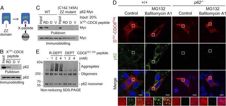
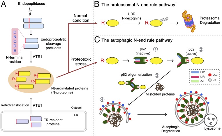
The conjugation of amino acids to the protein N termini is universally observed in eukaryotes and prokaryotes, yet its functions remain poorly understood. In eukaryotes, the amino acid l-arginine (l-Arg) is conjugated to N-terminal Asp (Nt-Asp), Glu, Gln, Asn, and Cys, directly or associated with posttranslational modifications. Following Nt-arginylation, the Nt-Arg is recognized by UBR boxes of N-recognins such as UBR1, UBR2, UBR4/p600, and UBR5/EDD, leading to substrate ubiquitination and proteasomal degradation via the N-end rule pathway. It has been a mystery, however, why studies for the past five decades identified only a handful of Nt-arginylated substrates in mammals, although five of 20 principal amino acids are eligible for arginylation. Here, we show that the Nt-Arg functions as a bimodal degron that directs substrates to either the ubiquitin (Ub)-proteasome system (UPS) or macroautophagy depending on physiological states. In normal conditions, the arginylated forms of proteolytic cleavage products, D101-CDC6 and D1156-BRCA1, are targeted to UBR box-containing N-recognins and degraded by the proteasome. However, when proteostasis by the UPS is perturbed, their Nt-Arg redirects these otherwise cellular wastes to macroautophagy through its binding to the ZZ domain of the autophagic adaptor p62/STQSM/Sequestosome-1. Upon binding to the Nt-Arg, p62 acts as an autophagic N-recognin that undergoes self-polymerization, facilitating cargo collection and lysosomal degradation of p62-cargo complexes. A chemical mimic of Nt-Arg redirects Ub-conjugated substrates from the UPS to macroautophagy and promotes their lysosomal degradation. Our results suggest that the Nt-Arg proteome of arginylated proteins contributes to reprogramming global proteolytic flux under stresses.

Keywords: ATE1 R-transferase; N-end rule pathway; macroautophagy; p62/STQSM/Sequestosome-1; ubiquitin-proteasome system.


Conflict of interest statement

Conflict of interest statement: Y.T.K., D.C.R., and Z.E. are coauthors on a 2016 Autophagy Guidelines publication that lists more than 1,000 authors who published their work in Autophagy.

Figures

Fig. 1.
Fig. 1.
Arginylated CDC6101–562 is normally degraded via the UPS-linked N-end rule pathway. (A) Sequence alignment of caspase 3-binding motifs of CDC6 proteins. (B) HeLa cells expressing X101-CDC6 (X = D, V, or RD) were treated with 10 μM MG132 for 5 h, followed by immunoblotting. (C) Cycloheximide degradation assay. HeLa cells expressing X101-CDC6-Flag were treated with 10 μM cycloheximide (CHX). (D and E) Same assay as B in +/+ and ATE1−/− MEFs. The GAPDH panel in E is identical to that in D. CDC6 was detected using anti-Flag in D and anti–R-CDC6 in E. (F) CHX degradation assay in +/+ and ATE1−/− MEFs.
Fig. 2.
Fig. 2.
Arginylated CDC6101–562 is degraded via autophagy under proteasomal inhibition. (A) HEK293 cells expressing RD101-CDC6-Flag were treated with 3 μM MG132 and 25 μM HCQ for 17 h. (B) MEFs expressing D101-CDC6-Flag were treated with 0.3 μM MG132 for 17 h alone or together with 0.2 μM BAF for 5 h, followed by anti-Flag immunostaining. (Scale bars, 10 μm.) (C) Same as B, followed by immunostaining of p62, LC3, or LAMP1. (Scale bars, 10 μm.) (D) Wild-type and ATG5−/− MEFs expressing X101-CDC6-Flag (X = V or RD) were subjected to immunoblotting analysis. (E) Wild-type and ATG5−/− MEFs expressing RD101-CDC6-Flag were treated with 1 μM cycloheximide (CHX) alone or together with 10 μM MG132, followed by immunoblotting. (F) HeLa cells were treated with MG132 alone or together with thapsigargin. (G) HeLa cells were treated with 3 μM MG132 singly or in combination with 0.2 μM BAF, 25 μM HCQ, and/or 0.2 μM thapsigargin (TG) for 17 h, followed by immunoblotting.
Fig. 3.
Fig. 3.
Arginylated CDC6101–562 binds and activates p62. (A) Diagram illustrating X-peptide pulldown assay using synthetic peptides. (B) HEK293 cell lysates were mixed with X101-CDC6 peptides (X = D, V, or RD), followed by immunoblotting analysis. (C) HEK293 cells expressing wild-type or C142/145A p62-myc were subjected to pulldown assay with X101-CDC6 peptides. (D) Wild-type and p62−/− MEFs expressing D101-CDC6-Flag were treated with 0.3 μM MG132 for 12 h, followed by cotreatment with 0.2 μM BAF for 5 h, and analyzed using anti-Flag immunostaining in comparison to p62. (Scale bars, 10 μm.) (E) HEK293 cell lysates were incubated with the tetrapeptide R-DEPT101–104 or DEPT101–104 for 2 h, followed by nonreducing SDS/PAGE.
Fig. 4.
Fig. 4.
P62 is a general receptor of cellular Nt-arginylated proteins. (A) HeLa cells expressing X1156-BRCA1-Flag (X = D, V, or RD) were treated with 10 μM MG132 for 5 h and immunoblotted. (B) HeLa cells expressing X1156-BRCA1-Flag were treated with 10 μM cycloheximide (CHX), followed by immunoblotting. Relative band intensities were normalized to those with no treatment. (C) MEFs were treated with 0.3 μM MG132 12 h, followed by cotreatment with 0.2 μM BAF for 5 h, and analyzed using anti-Flag immunostaining in comparison to p62, LC3, and LAMP1. (Scale bars, 10 μm.) (D) HEK293 cell lysates were subjected to X-peptide pulldown assay of p62 using X-BRCA11156–1166 peptides. (E) Sequences of N-terminal regions of (putative) arginylation substrates used in F. (F) X-peptides were mixed with HEK293 cell extracts to pulldown p62. IB, immunoblotting.
Fig. 5.
Fig. 5.
Nt-arginylation modulates for autophagic proteolysis. (A) HEK293 cells expressing X101-CDC6-Flag were treated with 1 μM geldanamycin and 25 μM HCQ for 17 h. (B) HeLa cells were treated with 3 μM MG132 alone or together with 0.2 μM thapsigargin. (C) Control and ATE1−/− MEFs were treated with 0.3 μM MG132 alone for 17 h or, alternatively, for 12 h, followed by cotreatment with 0.2 μM BAF for 5 h. (D) Control and ATE1−/− MEFs were treated with 0.3 μM MG132 and subjected to immunoblotting, followed by immunoblotting analysis using FK2 antibody specific to Ub conjugates. (E) Pregnant ATE1+/− female mice were injected i.v. with 20 mg/kg MG132 for 3 d. The resulting embryos and littermate controls at E12.5 were harvested, and their gross morphology was examined. (Scale bars, 1 mm.)
Fig. 6.
Fig. 6.
Chemical mimicry of Nt-Arg activates autophagy and facilitates autophagic targeting of misfolded proteins. (A) C57BL/6 mice were injected i.v. with 10 mg/kg XIE62-1004. Brains were harvested 24 h after treatment, paraffin-sectioned, and immunostained for LC3 and p62. Shown are cortex areas. (Scale bars, 10 μm.) (B) HeLa cells were treated with 3 μM MG132 alone or together with 10 μM XIE62-1004 for indicated times, followed by immunoblotting analysis of Ub conjugates. (C) HeLa cells were treated with 3 μM MG132 alone or together with 10 μM XIE62-1004 for 12 h, followed by cotreatment with 0.2 μM BAF for 5 h and immunostaining with FK2 and LC3 antibodies. (Scale bars, 10 μm.) (D) HeLa cells expressing CL1-YFP were treated with 25 μM HCQ alone or in combination with 10 μM XIE62-1004 for 17 h, followed by immunostaining analysis of LC3. (Scale bars, 10 μm.) (E) HeLa cells were incubated with 25 μM X-TAT-FITC (X = R or V) peptide for 18 h, followed by FITC fluorescence analysis. (Scale bars, 10 μm.) (F) HeLa cells were incubated with native TAT-FITC or X-TAT-FITC (X = R or V) for 8 h, followed by cotreatment with 0.1 μM BAF for 4 h and immunostaining analysis of LC3 in comparison to FITC fluorescence. (Scale bars, 10 μm.)
Fig. 7.
Fig. 7.
Hypothetical model illustrating the role of Nt-arginylation as a bimodal degron that modulates the cross-talk between the UPS and autophagy. (A) In eukaryotic cells, large numbers of cleavage products are generated by exopeptidases and endopeptidases. (B) Some of them are Nt-arginylated, and thus are normally degraded through ubiquitination via the UPS-linked N-end rule pathway. (C) However, when Nt-arginylated substrates cannot be properly processed by the proteasome under cellular stresses, they are metabolically stabilized, possibly associated with reduced activities of N-end rule machinery, resulting in increased concentrations of the Nt-Arg. The Nt-Arg of arginylated substrates from various subcellular compartments, such as the cytosol and ER, forms an N-proteome that directly binds the ZZ domain of p62 (step 1). The binding induces self-polymerization of p62 through a conformational change from a closed configuration to an open configuration (step 2), facilitating the collection of cargoes such as misfolded proteins and their aggregates (step 3) and, thus accelerating the delivery of p62–cargo complexes to autophagosomes (step 4). Through this mechanism, cells can sense and react to accumulating autophagic cargoes by activating p62 in a timely fashion using otherwise protein “wastes.”

References

    1. Ciechanover A, Kwon YT. Degradation of misfolded proteins in neurodegenerative diseases: Therapeutic targets and strategies. Exp Mol Med. 2015;47:e147. - PMC - PubMed
    1. Ciechanover A, Kwon YT. Protein quality control by molecular chaperones in neurodegeneration. Front Neurosci. 2017;11:185. - PMC - PubMed
    1. Deng Z, et al. Autophagy receptors and neurodegenerative diseases. Trends Cell Biol. 2017;27:491–504. - PubMed
    1. Cha-Molstad H, et al. Amino-terminal arginylation targets endoplasmic reticulum chaperone BiP for autophagy through p62 binding. Nat Cell Biol. 2015;17:917–929. - PMC - PubMed
    1. Tasaki T, Sriram SM, Park KS, Kwon YT. The N-end rule pathway. Annu Rev Biochem. 2012;81:261–289. - PMC - PubMed

Publication types

MeSH terms