. 2018 Mar 6;8(1):60.
doi: 10.1038/s41398-018-0106-x.

Fear extinction requires infralimbic cortex projections to the basolateral amygdala

Affiliations

Fear extinction requires infralimbic cortex projections to the basolateral amygdala

Daniel W Bloodgood et al. Transl Psychiatry. .

Abstract

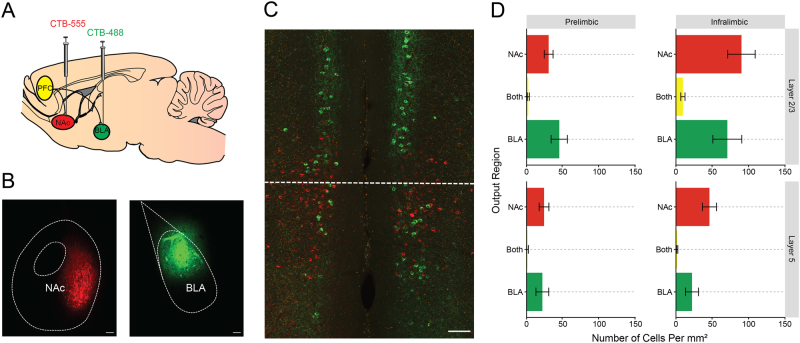
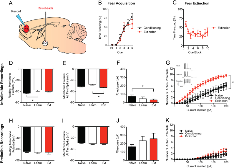
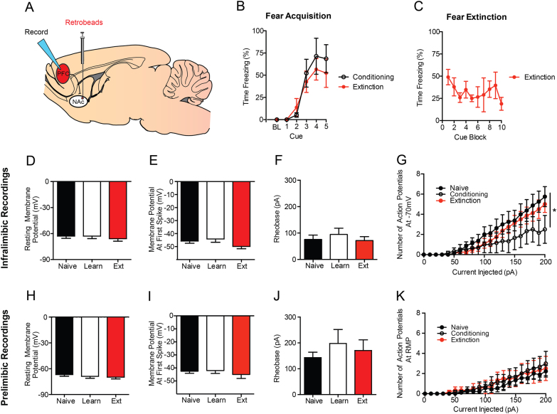
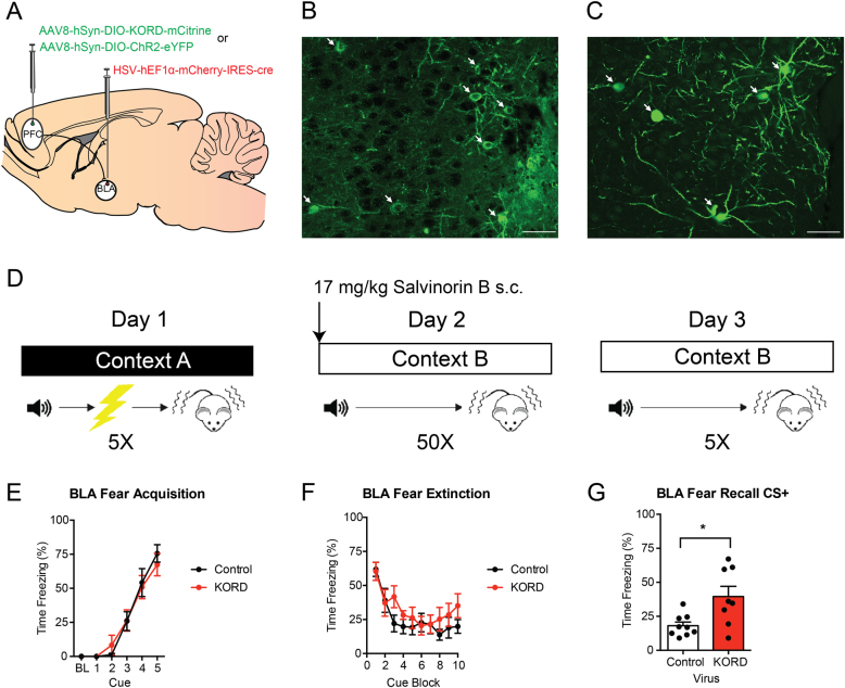
Fear extinction involves the formation of a new memory trace that attenuates fear responses to a conditioned aversive memory, and extinction impairments are implicated in trauma- and stress-related disorders. Previous studies in rodents have found that the infralimbic prefrontal cortex (IL) and its glutamatergic projections to the basolateral amygdala (BLA) and basomedial amygdala (BMA) instruct the formation of fear extinction memories. However, it is unclear whether these pathways are exclusively involved in extinction, or whether other major targets of the IL, such as the nucleus accumbens (NAc) also play a role. To address this outstanding issue, the current study employed a combination of electrophysiological and chemogenetic approaches in mice to interrogate the role of IL-BLA and IL-NAc pathways in extinction. Specifically, we used patch-clamp electrophysiology coupled with retrograde tracing to examine changes in neuronal activity of the IL and prelimbic cortex (PL) projections to both the BLA and NAc following fear extinction. We found that extinction produced a significant increase in the intrinsic excitability of IL-BLA projection neurons, while extinction appeared to reverse fear-induced changes in IL-NAc projection neurons. To establish a causal counterpart to these observations, we then used a pathway-specific Designer Receptors Exclusively Activated by Designer Drugs (DREADD) strategy to selectively inhibit PFC-BLA projection neurons during extinction acquisition. Using this approach, we found that DREADD-mediated inhibition of PFC-BLA neurons during extinction acquisition impaired subsequent extinction retrieval. Taken together, our findings provide further evidence for a critical contribution of the IL-BLA neural circuit to fear extinction.


Conflict of interest statement

Dr. Sugam is currently employed by Merck & Co., Inc. (Kenilworth, NJ, USA) however all data collection and analysis was performed at UNC prior to employment with Merck. The remaining authors declare that no conflict of interests.

Figures

Fig. 1
Fig. 1. Distinct mPFC projections to the BLA and NAc.
a The retrograde tracer CTB separately labeled with Alexa 488 and Alexa 555 was injected into the NAc and BLA. b Representative image of injection sites in the BLA and NAc. c Injection of tracers led to dense, but non-overlapping labeling of cells in the IL and PL. d Quantification of fluorescent cells revealed dense, non-overlapping labeling of BLA and NAc outputs throughout the PFC that was concentrated in the superficial layers of IL. Scale bar represents 100 µm. All the data presented are mean ± SEM. n = 18 images from N = 6 mice
Fig. 2
Fig. 2. Extinction increases neuronal excitability in the IL-BLA, but not PL-BLA, pathway.
a Retrobeads were injected into the BLA to visualize IL-BLA and PL-BLA projection neurons for electrophysiology recordings. b Fear conditioning increased freezing across CS-US pairings. c Fear extinction decreased freezing across CS trial-blocks. In IL-BLA projection neurons, fear conditioning resulted in (d) increased resting membrane potential that was not present after extinction. In IL-BLA neurons, following extinction training there was also (e) higher action potential threshold, (f) lower rheobase, and (g) an increase in the number of action potentials fired across increasing current injections. (Inset) Representative traces of current-injected firing during the final step of an increasing current injection protocol. In PL-BLA projections neurons, there were no differences in (h) resting membrane potential, (i) action potential threshold, (j) rheobase, or (k) action potential number with increasing current injection magnitude. The data presented are tests completed at resting membrane potential. All the data presented are mean ± SEM. The data were collected from N = 6 mice per group. In the infralimbic cortex there were n = 8 cells per experiment in the naive group, n = 8 cells in the fear learning group, and n = 10 cells in the fear extinction group. In the prelimbic cortex, there were n = 8 cells per experiment in the naive group, n = 5 cells in the fear learning group, and n = 4 cells in the fear extinction group. “*” denotes p < 0.05
Fig. 3
Fig. 3. Extinction does not increase neuronal excitability in the IL-NAc or PL-NAc pathways.
a Retrobeads were injected into the NAc to visualize IL-NAc projection neurons for electrophysiology recordings. b Fear conditioning increased freezing across CS-US pairings. c Fear extinction decreased freezing across CS trial-block. In IL-NAc projection neurons, there were no differences following fear conditioning or fear extinction in (h) resting membrane potential, (i) action potential threshold, or (j) rheobase. (g) There was a significant reduction in the number of action potentials fired in the fear conditioning group when recordings were performed at −70 mV, but was not present when performed at resting membrane potential. In PL-NAc projections neurons, there were no differences in (h) resting membrane potential, (i) action potential threshold, (j) rheobase, or (k) action potential number with increasing current injection magnitude. All the data presented are mean ± SEM. The data were collected from N = 4 mice per group. In the infralimbic cortex, there were n = 8 cells per experiment in the naive group, n = 6 cells in the fear learning group, and n = 8 cells in the fear extinction group. In the prelimbic cortex, there were n = 9 cells per experiment in the naive group, n = 6 cells in the fear learning group, and n = 6 cells in the fear extinction group. * denotes p < 0.05
Fig. 4
Fig. 4. Chemogenetic inhibition of IL-BLA projections impairs long-term extinction memory formation.
a HSV was injected into the BLA and KORD or a control virus was injected into the IL to selectively inhibit IL-BLA projections. Representative images of (b) KORD and (c) eYFP expression in the IL. d Mice were fear conditioned on day 1, then injected with Sal-B prior to extinction acquisition on day 2 and tested, drug-free, for extinction retrieval on day 3. Bottom row, PFC-BLA behavior cohort: (e) Fear conditioning increased freezing across CS-US pairings. f Fear extinction decreased freezing across CS presentations, irrespective of IL-BLA inhibition. g IL-BLA inhibition during extinction acquisition impaired extinction memory retrieval. All data presented are mean ± SEM. N = 8 KORD mice and N = 10 controls. * denotes p < 0.05

References

    1. Milad MR, Quirk GJ. Fear extinction as a model for translational neuroscience: ten years of progress. Annu. Rev. Psychol. 2012;63:129–151. doi: 10.1146/annurev.psych.121208.131631. - DOI - PMC - PubMed
    1. Bukalo O, Pinard CR, Holmes A. Mechanisms to medicines: elucidating neural and molecular substrates of fear extinction to identify novel treatments for anxiety disorders. Br. J. Pharmacol. 2014;171:4690–4718. doi: 10.1111/bph.12779. - DOI - PMC - PubMed
    1. Singewald N, Schmuckermair C, Whittle N, Holmes A, Ressler KJ. Pharmacology of cognitive enhancers for exposure-based therapy of fear, anxiety and trauma-related disorders. Pharmacol. Ther. 2015;149:150–190. doi: 10.1016/j.pharmthera.2014.12.004. - DOI - PMC - PubMed
    1. Tovote P, Fadok JP, Luthi A. Neuronal circuits for fear and anxiety. Nat. Rev. Neurosci. 2015;16:317–331. doi: 10.1038/nrn3945. - DOI - PubMed
    1. Rozeske RR, Valerio S, Chaudun F, Herry C. Prefrontal neuronal circuits of contextual fear conditioning. Genes. Brain. Behav. 2015;14:22–36. doi: 10.1111/gbb.12181. - DOI - PubMed