

Observation of optic disc neovascularization using OCT angiography in proliferative diabetic retinopathy after intravitreal conbercept injections
- PMID: 29507304
- PMCID: PMC5838089
- DOI: 10.1038/s41598-018-22363-0
Observation of optic disc neovascularization using OCT angiography in proliferative diabetic retinopathy after intravitreal conbercept injections
Abstract
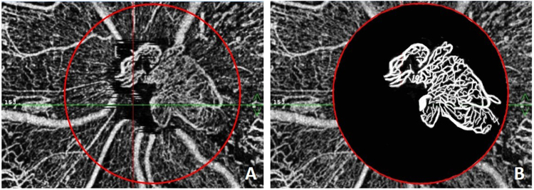
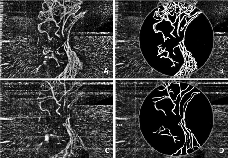
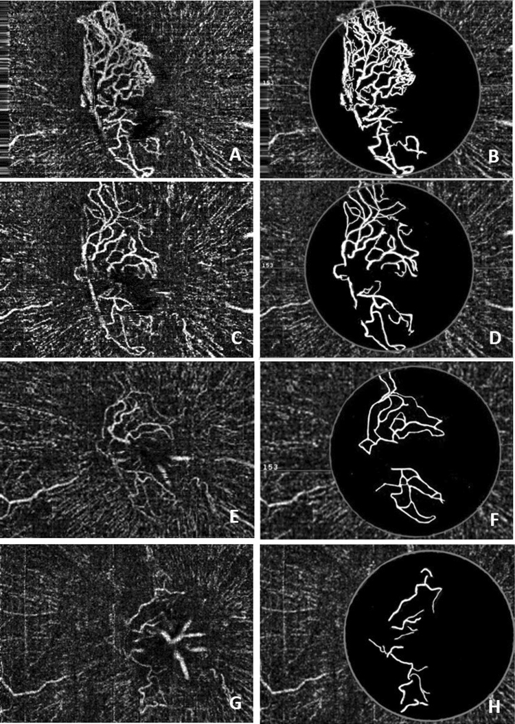
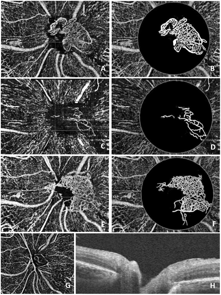
This study reports the short-term efficacy and safety of intravitreal conbercept injections for neovascularization at the disc (NVD) in patients with proliferative diabetic retinopathy (PDR). Conbercept is a recombinant fusion protein with a high affinity for all isoforms of vascular endothelial growth factor (VEGF)-A, placental growth factor and VEGF-B. A prospective case series study was conducted in 15 patients (15 eyes). Patients had complete ocular examinations and received a 0.5 mg intravitreal conbercept injection followed by supplemental pan-retinal photocoagulation (PRP). Optical coherence tomography angiography (OCTA) was performed before and after treatment. Before treatment, the mean NVD area was 1.05 ± 0.33 mm2, and it decreased to 0.56 ± 0.17 mm2 after an interval of 7.5 d (p = 0.000). One eye required vitrectomy during follow-up. Recurrent NVD was observed in 2 eyes, which resolved after repeated injections. The remaining 12 eyes were stable over a mean follow-up period of 8.3 months. The mean area of the NVD in 14 patients without vitrectomy was 0.22 ± 0.11 mm2 (p = 0.000) at the last visit. Intravitreal conbercept injections combined with intensive PRP are an effective and safe treatment for PDR with NVD. Quantitative information on NVD can be obtained with OCTA, which may be clinically useful in evaluating the therapeutic effect.
Conflict of interest statement
The authors declare no competing interests.
Figures
Similar articles
-
Intravitreal bevacizumab (Avastin) and panretinal photocoagulation in the treatment of high-risk proliferative diabetic retinopathy.J Ocul Pharmacol Ther. 2013 Jul-Aug;29(6):550-5. doi: 10.1089/jop.2012.0202. Epub 2013 Mar 15. J Ocul Pharmacol Ther. 2013. PMID: 23495932 Free PMC article. Clinical Trial.
-
Comparison of aqueous humor levels of PlGF and VEGF in proliferative diabetic retinopathy before and after intravitreal conbercept injection.Diabetes Res Clin Pract. 2020 Apr;162:108083. doi: 10.1016/j.diabres.2020.108083. Epub 2020 Feb 11. Diabetes Res Clin Pract. 2020. PMID: 32057965
-
Vascular endothelial growth factor concentration in vitreous humor of patients with severe proliferative diabetic retinopathy after intravitreal injection of conbercept as an adjunctive therapy for vitrectomy.Chin Med J (Engl). 2020 Mar 20;133(6):664-669. doi: 10.1097/CM9.0000000000000687. Chin Med J (Engl). 2020. PMID: 32068603 Free PMC article. Clinical Trial.
-
Intravitreal conbercept improves outcome in patients undergoing vitrectomy for proliferative diabetic retinopathy: A systematic review and meta-analysis.J Evid Based Med. 2020 May;13(2):116-124. doi: 10.1111/jebm.12379. Epub 2020 Mar 13. J Evid Based Med. 2020. PMID: 32167242
-
New Findings in Diabetic Maculopathy and Proliferative Disease by Swept-Source Optical Coherence Tomography Angiography.Dev Ophthalmol. 2016;56:113-21. doi: 10.1159/000442802. Epub 2016 Mar 15. Dev Ophthalmol. 2016. PMID: 27023703 Review.
Cited by
-
Optical Coherence Tomography Angiography Features of Neovascularization in Proliferative Diabetic Retinopathy.Clin Ophthalmol. 2020 Oct 15;14:3351-3362. doi: 10.2147/OPTH.S274537. eCollection 2020. Clin Ophthalmol. 2020. PMID: 33116386 Free PMC article.
-
Optical coherence tomography features of neovascularization in proliferative diabetic retinopathy: a systematic review.Int J Retina Vitreous. 2020 Jun 29;6:26. doi: 10.1186/s40942-020-00230-3. eCollection 2020. Int J Retina Vitreous. 2020. PMID: 32612851 Free PMC article. Review.
-
Longitudinal Wide-Field Swept-Source OCT Angiography of Neovascularization in Proliferative Diabetic Retinopathy after Panretinal Photocoagulation.Ophthalmol Retina. 2019 Apr;3(4):350-361. doi: 10.1016/j.oret.2018.11.008. Epub 2018 Nov 24. Ophthalmol Retina. 2019. PMID: 31014688 Free PMC article.
-
Swept-source optical coherence tomography angiography of diabetic papillopathy: a case report.BMC Ophthalmol. 2020 May 15;20(1):194. doi: 10.1186/s12886-020-01470-5. BMC Ophthalmol. 2020. PMID: 32414351 Free PMC article.
-
Interplay of endothelial-mesenchymal transition, inflammation, and autophagy in proliferative diabetic retinopathy pathogenesis.Heliyon. 2024 Feb 1;10(3):e25166. doi: 10.1016/j.heliyon.2024.e25166. eCollection 2024 Feb 15. Heliyon. 2024. PMID: 38327444 Free PMC article.
References
-
- Early Treatment Diabetic Retinopathy Study Research Group Early photocoagulation for diabetic retinopathy. ETDRS report number 9. Ophthalmology. 1991;98(5 suppl):766–785. - PubMed
-
- Avery RL, et al. Intravitreal bevacizumab (Avastin) in the treatment of proliferative diabetic retinopathy. Ophthalmology. 2006;113:1695.e1–15. - PubMed
MeSH terms
Substances
LinkOut - more resources
Full Text Sources
Other Literature Sources
Medical
Research Materials
