. 2018 Apr;10(4):420-427.
doi: 10.1038/s41557-018-0008-9. Epub 2018 Mar 5.

Evolution of sequence-defined highly functionalized nucleic acid polymers

Affiliations

Evolution of sequence-defined highly functionalized nucleic acid polymers

Zhen Chen et al. Nat Chem. 2018 Apr.

Abstract

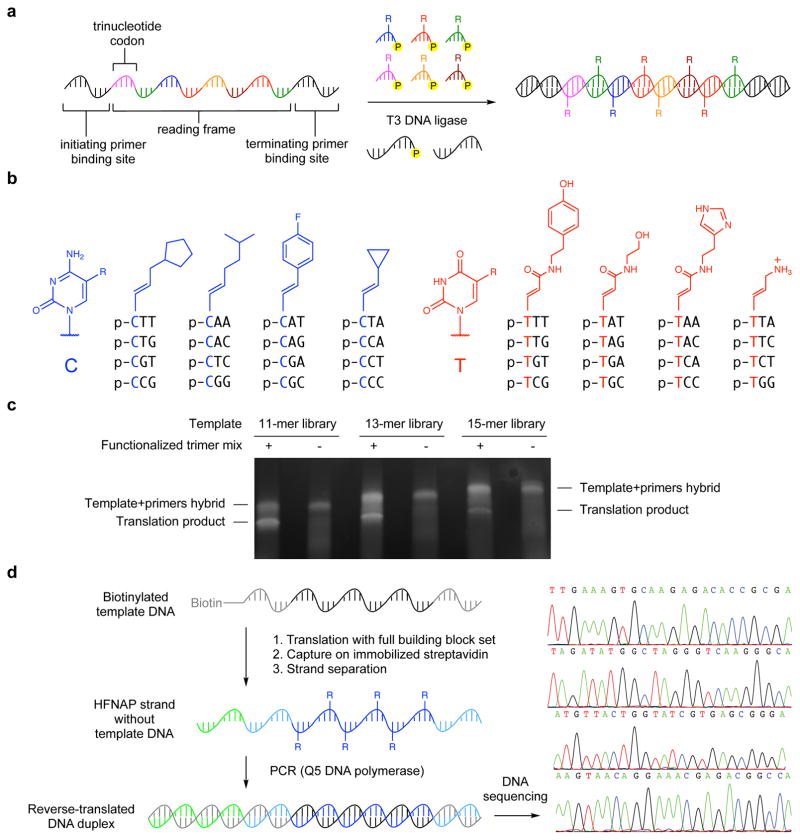
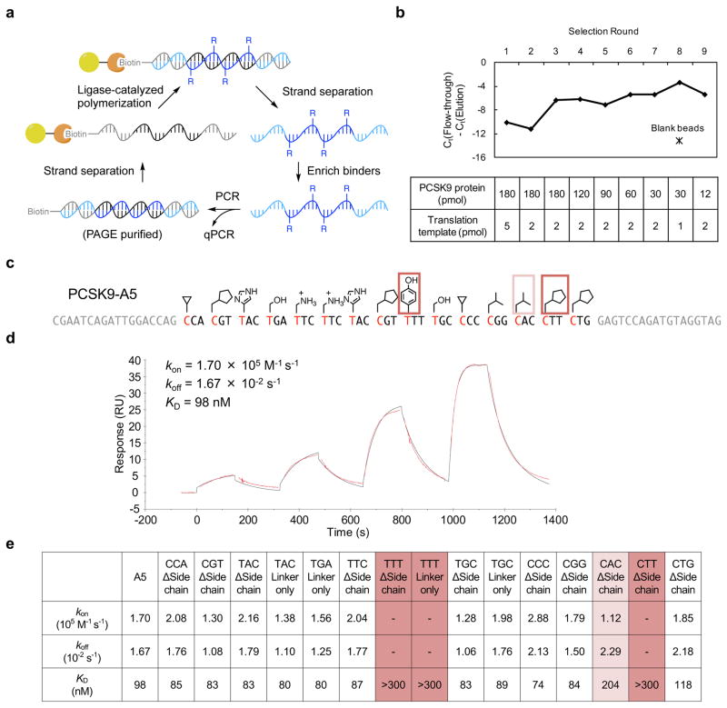
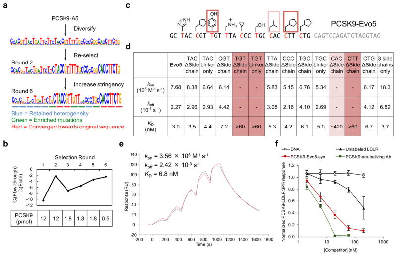
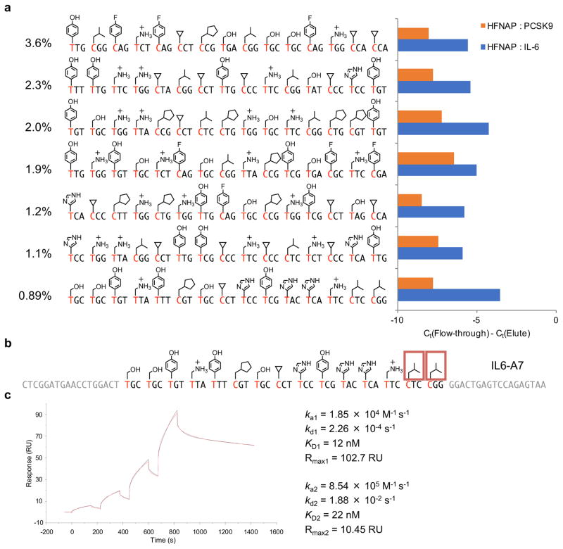
The evolution of sequence-defined synthetic polymers made of building blocks beyond those compatible with polymerase enzymes or the ribosome has the potential to generate new classes of receptors, catalysts and materials. Here we describe a ligase-mediated DNA-templated polymerization and in vitro selection system to evolve highly functionalized nucleic acid polymers (HFNAPs) made from 32 building blocks that contain eight chemically diverse side chains on a DNA backbone. Through iterated cycles of polymer translation, selection and reverse translation, we discovered HFNAPs that bind proprotein convertase subtilisin/kexin type 9 (PCSK9) and interleukin-6, two protein targets implicated in human diseases. Mutation and reselection of an active PCSK9-binding polymer yielded evolved polymers with high affinity (KD = 3 nM). This evolved polymer potently inhibited the binding between PCSK9 and the low-density lipoprotein receptor. Structure-activity relationship studies revealed that specific side chains at defined positions in the polymers are required for binding to their respective targets. Our findings expand the chemical space of evolvable polymers to include densely functionalized nucleic acids with diverse, researcher-defined chemical repertoires.


Conflict of interest statement

Competing Financial Interests

The authors have filed patent applications on aspects of this work.

Figures

Figure 1
Figure 1. Design and construction of the sequence-defined polymer library
a, Reaction scheme for DNA ligase-mediated translation of DNA templates into sequence-defined highly functionalized nucleic acid polymers (HFNAPs). b, Structures of 5′-phosphorylated trinucleotide building blocks for HFNAP library synthesis. c, Translation of libraries of randomized DNA templates into HFNAPs that incorporate up to 15 consecutive functionalized trinucleotide building blocks. The translation reactions, as well as control reactions from which the trinucleotide building blocks were omitted, were analyzed by polyacrylamide gel electrophoresis on a non-denaturing 10% TBE gel and imaged by SYBR gold staining. d, A complete cycle of HFNAP translation, HFNAP strand isolation, and reverse translation back into DNA faithfully recovered sequence information from the original DNA templates. Left: experimental scheme; right: electrophoretic traces from Sanger sequencing. In control experiments in which the trinucleotide building blocks were omitted from the polymerization reactions, the PCR step did not generate any amplicons of the correct size.
Figure 2
Figure 2. Selection of PCSK9-binding polymers from a random HFNAP library
a, Overview of translation, selection, and reverse translation scheme. b, PCSK9 binding selection progress. The HFNAP pool’s bulk affinity to PCSK9-coated beads was assessed by quantifying the amount of HFNAP in the flow-through versus the elution at each round of selection by quantitative PCR. Higher values in the graph indicate higher ratios of polymers that bound to immobilized PCSK9 and were eluted relative to polymers that flowed through the immobilized PCSK9. c, Sequence and side-chain structure of selected polymer PCSK9-A5. Side-chains essential for binding activity are boxed. d, SPR sensogram characterizing binding kinetics between surface-immobilized PCSK9-A5 polymer and the target PCSK9 protein. The concentrations of injected PCSK9 were 10, 30, 100, and 300 nM. The observed sensogram is shown in red and the fitted curve with the kinetic parameters listed is shown in black. e, Kinetic parameters for binding of PCSK9-A5 or its side-chain-deficient variants to PCSK9 protein, as measured by SPR. For the variants “TTT ΔSide chain”, “TTT Linker only”, and “CTT ΔSide chain”, no SPR signal was observed at highest analyte concentration tested (300 nM PCSK9).
Figure 3
Figure 3. Evolution of an improved PCSK9-binding polymer
a, Evolution scheme and DNA sequencing results of the diversification and iterated selection of PCSK9-A5 variants with increased PCSK9 binding activity. b, Affinity maturation of the diversified PCSK9-A5 pool. The evolving polymer pool’s bulk affinity to immobilized PCSK9 was assessed by quantifying the amount of HFNAP in the flow-through and the elution at each round of selection by quantitative PCR. c, Sequence and side-chain structure of the resulting PCSK9-Evo5 polymer. Side chains essential for binding activity are boxed. d, Kinetic parameters for binding of PCSK9-Evo5 or its side-chain-deficient variants to PCSK9 protein, as measured by SPR. For the variants “TGT ΔSide chain”, “TGT Linker only”, and “CTT ΔSide chain”, no SPR signal was observed at the highest analyte concentration tested (60 nM PCSK9). For the variant “CAC ΔSide chain”, the binding interaction fits a two-state reaction kinetic model with KD ≈ 420 nM. Representative sensograms are provided in Supplementary Fig. 5. e, SPR sensogram characterizing binding kinetics between PCSK9-Evo5-syn and surface-immobilized PCSK9 protein. The concentrations of injected PCSK9-Evo5-syn were 1.8, 6, 18, 60, and 180 nM. The observed sensogram is shown in red and the fitted curve with the kinetic parameters listed is shown in black. f, SPR response on an LDLR-coated surface produced by flowing PCSK9 in the presence of either PCSK9-Evo5-syn, unfunctionalized DNA of identical sequence to PCSK9-Evo5-syn, unlabeled LDLR, or a known PCSK9-neutralizing monoclonal antibody. The SPR response shown is normalized to the response in experiments without any competitor (defined as an SPR response of 1). Error bars represent s.d. (n = 3). Representative raw sensograms are provided in Supplementary Fig. 11.
Figure 4
Figure 4. Characterization of IL-6-binding HFNAPs selected from a random library
a, Retention of individual selection-enriched HFNAPs on immobilized IL-6 (target; blue bars) or immobilized PCSK9 (non-target; red bars). The percentages of each sequence in the pool after seven rounds of selection are listed to the left. The experiment was performed as a screen to identify hits for further characterization, and was not performed in replicates. b, Sequence and side-chain structure of IL6-A7. Side-chains essential for binding activity are boxed. c, SPR sensogram characterizing binding kinetics between biotinylated IL6-A7 and its target IL-6 protein. The concentrations of injected IL-6 were 10, 30, 100, and 300 nM. The observed sensogram is shown in red and the fitted curve with the kinetic parameters listed is shown in black.

References

    1. Ellington AD, Szostak JW. In vitro selection of RNA molecules that bind specific ligands. Nature. 1990;346:818–822. - PubMed
    1. Tuerk C, Gold L. Systematic evolution of ligands by exponential enrichment: RNA ligands to bacteriophage T4 DNA polymerase. Science. 1990;249:505–510. - PubMed
    1. Robertson DL, Joyce GF. Selection in vitro of an RNA enzyme that specifically cleaves single-stranded DNA. Nature. 1990;344:467–468. - PubMed
    1. Bock LC, Griffin LC, Latham JA, Vermaas EH, Toole JJ. Selection of single-stranded DNA molecules that bind and inhibit human thrombin. Nature. 1992;355:564–566. - PubMed
    1. Ellington AD, Szostak JW. Selection in vitro of single-stranded DNA molecules that fold into specific ligand-binding structures. Nature. 1992;355:850–852. - PubMed

Publication types