. 2018 Mar 5;8(1):4020.
doi: 10.1038/s41598-018-22156-5.

Neutrophil extracellular trap-microparticle complexes enhance thrombin generation via the intrinsic pathway of coagulation in mice

Affiliations

Neutrophil extracellular trap-microparticle complexes enhance thrombin generation via the intrinsic pathway of coagulation in mice

Yongzhi Wang et al. Sci Rep. .

Abstract

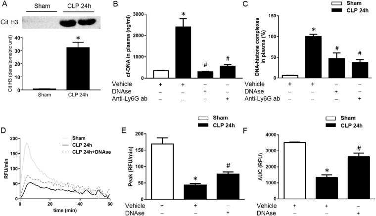
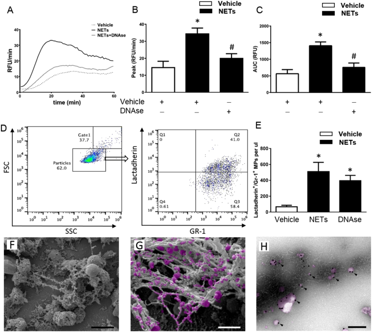
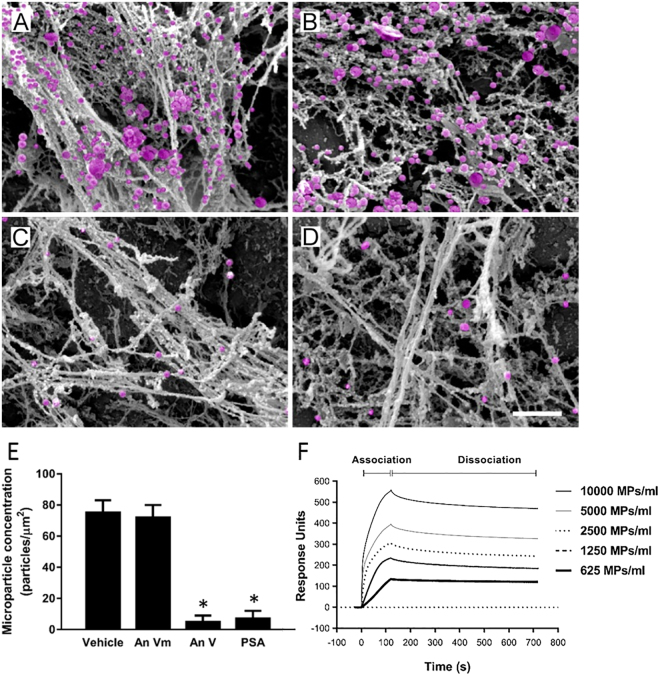
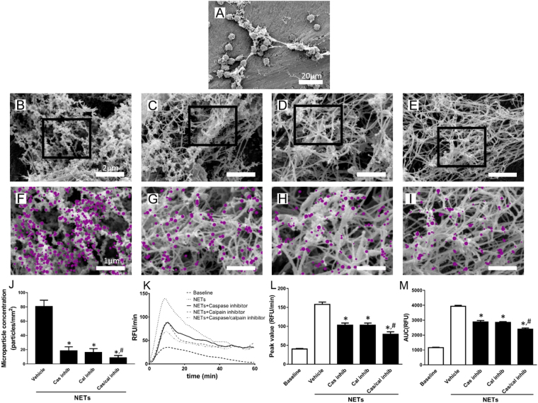
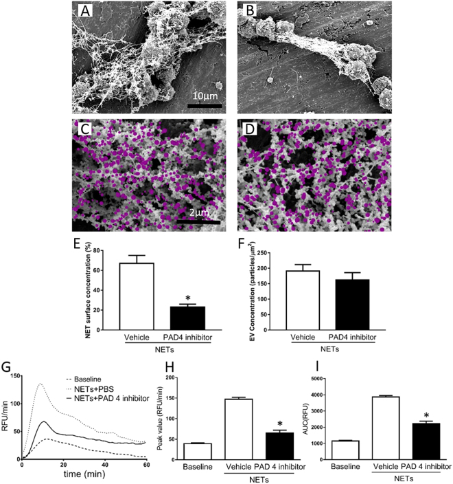
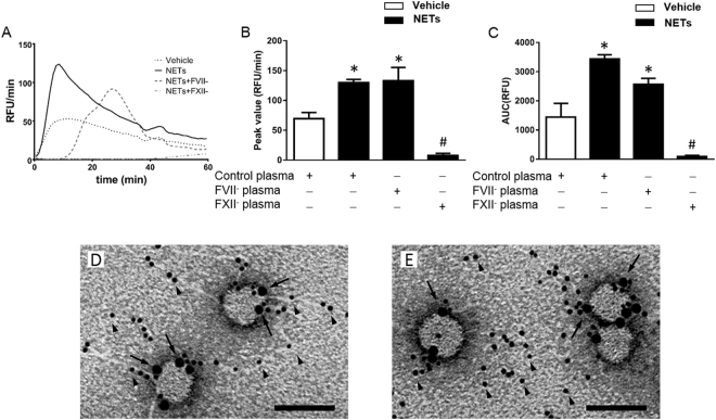
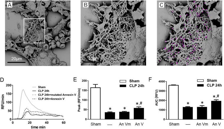
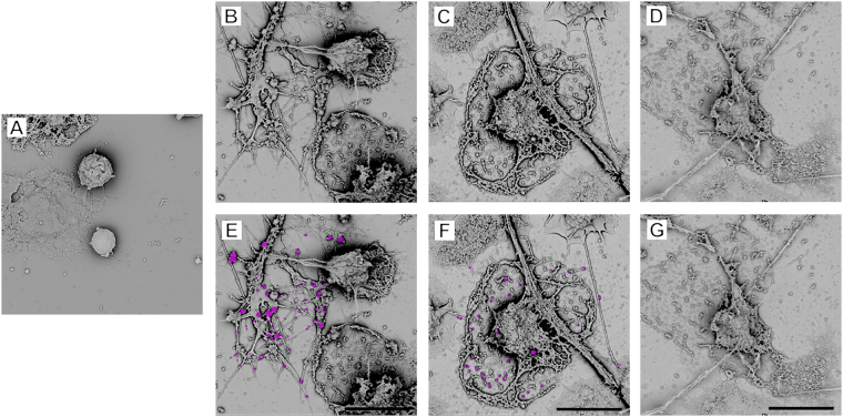
Abdominal sepsis is associated with dysfunctional hemostasis. Thrombin generation (TG) is a rate-limiting step in systemic coagulation. Neutrophils can expell neutrophil extracellular traps (NETs) and/or microparticles (MPs) although their role in pathological coagulation remains elusive. Cecal ligation and puncture (CLP)-induced TG in vivo was reflected by a reduced capacity of plasma from septic animals to generate thrombin. Depletion of neutrophils increased TG in plasma from CLP mice. Sepsis was associated with increased histone 3 citrullination in neutrophils and plasma levels of cell-free DNA and DNA-histone complexes and administration of DNAse not only eliminated NET formation but also elevated TG in sepsis. Isolated NETs increased TG and co-incubation with DNAse abolished NET-induced formation of thrombin. TG triggered by NETs was inhibited by blocking factor XII and abolished in factor XII-deficient plasma but intact in factor VII-deficient plasma. Activation of neutrophils simultaneously generated large amount of neutrophil-derived MPs, which were found to bind to NETs via histone-phosphatidylserine interactions. These findings show for the first time that NETs and MPs physically interact, and that NETs might constitute a functional assembly platform for MPs. We conclude that NET-MP complexes induce TG via the intrinsic pathway of coagulation and that neutrophil-derived MPs play a key role in NET-dependent coagulation.


Conflict of interest statement

The authors declare no competing interests.

Figures

Figure 1
Figure 1
NET formation in abdominal sepsis. (A) Representative western blot and aggregate data on citrullinated histone 3 in neutrophils isolated from the blood from sham and CLP animals. Plasma levels of (B) cf-DNA and (C) DNA-histone complexes were determined 24 h after CLP. (D) TG over time, (E) peak and (F) total levels of TG were determined as described in Material and Methods. Mice underwent CLP or the identical laparotomy and resuscitation procedures, but the cecum was neither ligated nor punctured (Sham). Animals received intraperitoneal injections of DNAse (5 mg/kg), an anti-Ly-6G antibody (Anti-Ly-6G ab, 20 mg/kg) or vehicle. Data are presented as mean ± SEM and n = 5. *P < 0.05 vs. Sham and #P < 0.05 vs. Vehicle + CLP.
Figure 2
Figure 2
NET-induced generation of thrombin. NETs were generated from PMA-stimulated bone marrow neutrophils. PPP was co-incubated with NETs containing vehicle or DNAse. (A) TG over time, (B) peak and (C) total levels of TG were determined as described in Material and Methods. Supernatants from PMA-stimulated neutrophils were incubated with FITC-conjugated bovine lactadherin and a PerCP Cy5.5-conjugated anti-Gr-1 antibody. (D) Particles were gated as events smaller than 1 μm in size and then gated as Lactadherin+/Gr-1+ and quantified by use of flow cytometry. (E) Aggregate data on NET-induced MPs. (F) Scanning electron microscopy showing neutrophils with extracellular web-like structures. Scale bar = 5 μm. (G) A higher magnification showing MPs attached to neutrophil-derived NETs. Scale bar = 2 μm. MPs are denoted in pink color. H) Transmission electron microscopy showing NETs and MPs incubated with gold-labeled annexin V (black particles, black arrowhead). Scale bar = 0.25 µm. Data are presented as mean ± SEM and n = 5. *P < 0.05 vs. Vehicle and #P < 0.05 vs. Vehicle + NETs.
Figure 3
Figure 3
MP binding to NETs. NETs were generated from PMA-stimulated bone marrow neutrophils co-incubated with (A) vehicle (B) mutated annexin V (An Vm), (C) wild-type annexin V (An V) or (D) polysialic acid (PSA). Scanning electron microscopy showing MPs attached to neutrophil-derived NETs. Scale bar = 2 μm. (E) Aggregate data on MP binding to NETs. (F) Surface plasmon resonance with histone H4 immobilized to a CM5 sensorchip. Indicated concentrations of MPs were applied in a flow over the sensorchip surface as described in Methods. Data are presented as mean ± SEM and n = 5. *P < 0.05 vs. Vehicle + CLP.
Figure 4
Figure 4
Functional role of NET-MP complexes. NETs were generated from PMA-stimulated bone marrow neutrophils (A). Scale bar = 20 µm. Then co-incubated with (B,F) vehicle (C,G) caspase inhibitor, (D,H) calpain inhibitor or (E,I) a combination of caspase and calpain inhibitors. Scanning electron microscopy showing MPs attached to neutrophil-derived NETs. Scale bar = 2 μm in the top insert and a higher magnification of the indicated area of interest from the top insert is shown below with a scale bar = 1 μm. MPs are denoted in pink color in the lower inserts. (J) Aggregate data on MP binding to NETs. (K) TG over time, (L) peak and (M) total levels of TG were determined as described in Material and Methods. Data are presented as mean ± SEM and n = 5. *P < 0.05 vs. Vehicle and #P < 0.05 vs. Vehicle + NETs.
Figure 5
Figure 5
Role of PAD4 in formation of NET-MP complexes. NETs were generated from PMA-stimulated bone marrow neutrophils co-incubated with (A,C) vehicle or (B,D) Cl-amidine, a PAD4 inhibitor. (A,B) Scanning electron microscopy showing MPs attached to neutrophil-derived NETs. Scale bar = 10μm and (C,D) a higher magnification with a scale bar = 2 μm. (C,D) MPs are denoted in pink color. Aggregate data on (E) NET formation and (F) MP per μm2 of of NETs. (G) TG over time, (H) peak and (I) total levels of TG were determined as described in Material and Methods. Data are presented as mean ± SEM and n = 5. *P < 0.05 vs. Vehicle + NETs.
Figure 6
Figure 6
NETs regulate TG via the intrinsic pathway of coagulation. NETs generated from PMA-stimulated bone marrow neutrophils were incubated in wild-type, factor VII- (FVII plasma) and factor XII-deficient (FXII plasma) PPP. (A) TG over time, (B) peak and (C) total levels of TG were determined as described in Material and Methods. Transmission electron microscopy showing NETs and MPs incubated with a gold-labeled anti-histone H4 antibody (small gold particles, black arrowhead) and (D) gold-labeled annexin V (large gold particles, black arrow) or (E) a gold-labeled anti-FXII antibody (large gold particles, black arrow). Scale bar = 0.25 µm. Data are presented as mean ± SEM and n = 5. *P < 0.05 vs. Vehicle + Control plasma and #P < 0.05 vs. NETs + Control plasma.
Figure 7
Figure 7
Sepsis-induced formation of NET-MP complexes. (A) Scanning electron microscopy showing extracellular web-like structures in the lung from a mouse exposed to CLP. Scale bar = 20 μm. (B) A higher magnification of the indicated area of interest from Figure A showing MPs attached to NETs in the septic lung. (C) MPs are denoted in pink color. Scale bar = 10 μm. (D) TG over time, (E) peak and (F) total levels of TG were determined as described in Material and Methods. Mice underwent CLP or the identical laparotomy and resuscitation procedures, but the cecum was neither ligated nor punctured (Sham). Animals received intraperitoneal injections of wild-type annexin V (An V) or mutated annexin V (An Vm). Data are presented as mean ± SEM and n = 5. *P < 0.05 vs. Sham and #P < 0.05 vs. An Vm + CLP.
Figure 8
Figure 8
NET-MP complex formation in human neutrophils. (A) Resting human neutrophils. NETs were generated from M1 protein-stimulated (2 h) human neutrophils co-incubated with (B,E) vehicle, (C,F) mutated annexin V and (D,G) wild-type annexin V. Scanning electron microscopy showing MPs attached to neutrophil-derived NETs. MPs are denoted in pink color in the lower inserts. Scale bar = 10 μm. One representative experiment of 5 independent experiments is shown.

Similar articles

Cited by

References

    1. Martin GS, Mannino DM, Eaton S, Moss M. The epidemiology of sepsis in the United States from 1979 through 2000. N Engl J Med. 2003;348:1546–1554. doi: 10.1056/NEJMoa022139. - DOI - PubMed
    1. Kaukonen KM, Bailey M, Suzuki S, Pilcher D, Bellomo R. Mortality related to severe sepsis and septic shock among critically ill patients in Australia and New Zealand, 2000–2012. JAMA. 2014;311:1308–1316. doi: 10.1001/jama.2014.2637. - DOI - PubMed
    1. Levi M, de Jonge E, van der Poll T. Sepsis and disseminated intravascular coagulation. J Thromb Thrombolysis. 2003;16:43–47. doi: 10.1023/B:THRO.0000014592.27892.11. - DOI - PubMed
    1. Levi M, van der Poll T. Inflammation and coagulation. Crit Care Med. 2010;38:S26–34. doi: 10.1097/CCM.0b013e3181c98d21. - DOI - PubMed
    1. Craciun FL, Schuller ER, Remick DG. Early enhanced local neutrophil recruitment in peritonitis-induced sepsis improves bacterial clearance and survival. J Immunol. 2010;185:6930–6938. doi: 10.4049/jimmunol.1002300. - DOI - PMC - PubMed

Publication types

LinkOut - more resources