. 2018 Feb 3;8(5):1411-1420.
doi: 10.7150/thno.22252. eCollection 2018.

Naphthalene-based fluorescent probes for glutathione and their applications in living cells and patients with sepsis

Affiliations

Naphthalene-based fluorescent probes for glutathione and their applications in living cells and patients with sepsis

Jun Li et al. Theranostics. .

Erratum in

Abstract

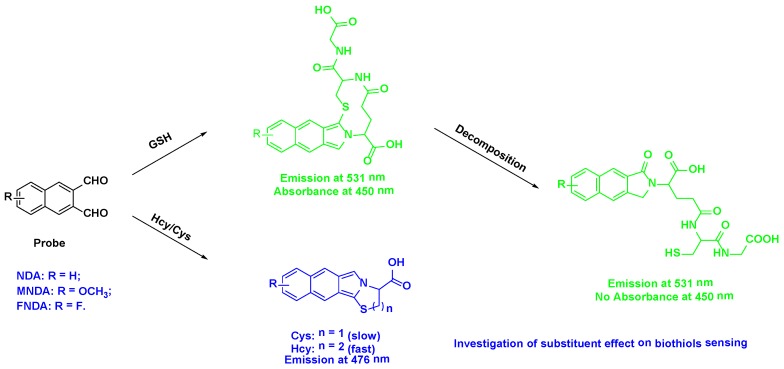
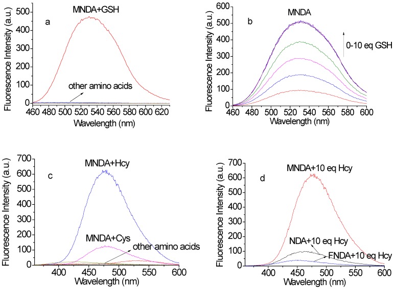
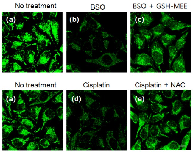
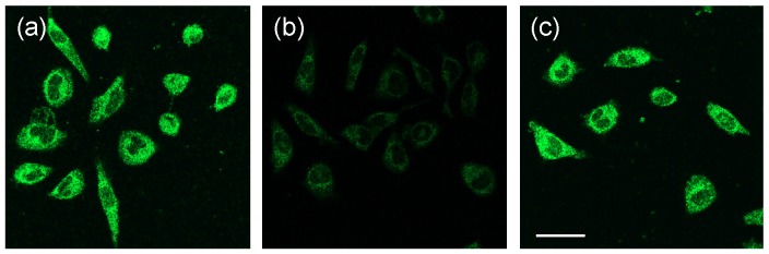
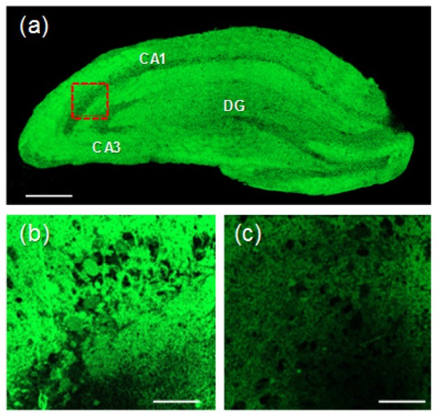
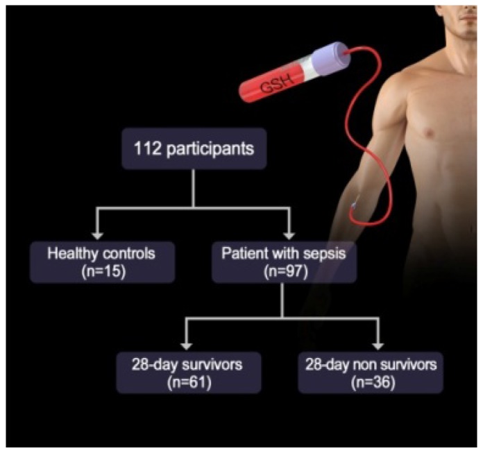
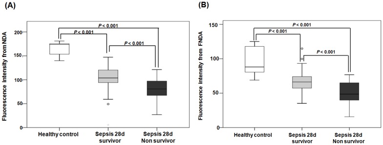
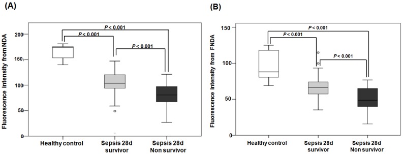
Rationale: Among the biothiols-related diseases, sepsis is defined as life-threatening organ dysfunction caused by a dysregulated host response to infection and can result in severe oxidative stress and damage to multiple organs. In this study, we aimed to develop a fluorescence chemosensor that can both detect GSH and further predict sepsis. Methods: In this study, two new naphthalene dialdehyde compounds containing different functional groups were synthesized, and the sensing abilities of these compounds towards biothiols and its applications for prediction of sepsis were investigated. Results: Our study revealed that the newly developed probe 6-methoxynaphthalene-2, 3-dicarbaldehyde (MNDA) has two-photon is capable of detecting GSH in live cells with two-photon microscopy (TPM) under the excitation at a wavelength of 900 nm. Furthermore, two GSH detection probes naphthalene-2,3-dicarboxaldehyde (NDA) and 6-fluoronaphthalene-2,3-dicarbaldehyde (FNDA) not only can detect GSH in living cells, but also showed clinical significance for the diagnosis and prediction of mortality in patients with sepsis. Conclusions: These results open up a promising direction for further medical diagnostic techniques.

Keywords: GSH; Sepsis; cell imaging; diagnosis; fluorescence probe.


Conflict of interest statement

Competing Interests: The authors have declared that no competing interest exists.

Figures

Figure 1
Figure 1
Simultaneous sensing of biothiols based on reactions with dialdehyde
Scheme 1
Scheme 1
Synthetic route of NDA derivatives. Reagents and conditions: (a) ethylene glycol, p-toluenesulfonic acid, toluene, reflux; (b) n-BuLi/DMF, THF, -78 oC, N2; (c) FeCl3.6H2O, acetone or 10% HCl, r.t; (d) PEt3, dimethylmaleate, DBU, CH2Cl2; (e) diisobutylaluminium hydride solution, -50 oC; (f) DMSO, (COCl)2, -78 oC, N2.
Figure 2
Figure 2
(a) Fluorescence responses of the newly developed probe MNDA (10 µM) to various amino acids in the green channel (including GSH, Hcy, Cys, Ala, DTT, Gln, Gly, His, Lys, Met, Pha and Ser, 100 µM each; excitation: 450 nm, slit widths: 3 nm, 3 nm). (b) Gradient titrations of MNDA with GSH in HEPES buffer solution. (c) Fluorescence responses of MNDA (10 µM) to various amino acids in the blue channel (including GSH, Hcy, Cys, Ala, DTT, Gln, Gly, His, Lys, Met, Pha and Ser, 100 µM each; excitation: 350 nm, slit widths: 1.5 nm, 3 nm); (d) Comparison of the sensing abilities of probes toward Hcy (excitation: 350 nm, slit widths: 1.5 nm, 3 nm).
Figure 3
Figure 3
Fluorescence imaging in cells. HeLa cells were pre-incubated with the following agents: (a) no treatment; (b) 1 mM NMM for 30 min; (c) 1 mM NMM for 30 min and 300 μM Cys for 30 min; (d) 1 mM NMM for 30 min and 300 µM Hcy for 30 min; (e) 1 mM NMM for 30 min and 1 mM GSH-MEE for 60 min. Cells were then washed with Dulbecco's phosphate-buffered saline (DPBS) and incubated with 10 µM MNDA for 30 min. After washing with DPBS, fluorescence images were acquired by confocal microscopy. Blue: ex. 405 nm/em. 430-455 nm, green: ex. 473 nm/em. 490-540 nm. Scale bar: 10 µm.
Figure 4
Figure 4
Changes in the intracellular GSH level after drug treatment. HeLa cells were incubated with (a) no treatment; (b) 100 µM BSO for 6 h; (c) 100 µM BSO with 1 mM GSH-MEE for 6 h; (d) 50 µM cisplatin for 6 h; (e) 50 µM cisplatin with 2 mM NAC for 6 h. Cells were then stained with 3 µM MNDA for 30 min and imaged withex. 473 nm/em. 490-590 nm. Scale bar: 10 µm.
Figure 5
Figure 5
TPM images of 10 µM MNDA-labelled HeLa cells. HeLa cells were pre-incubated with the following agents: (a) no treatment; (b) 1 mM NMM for 30 min; (c) 1 mM NMM for 30 min and 1 mM GSH-MEE for 60 min. Cells were then washed with Dulbecco's phosphate-buffered saline (DPBS) and incubated with 10 µM MNDA for 30 min. TPM images were excited at900 nm and acquired at 400-600 nm. Scale bar: 10 µm.
Figure 6
Figure 6
(a) Accumulated TPM images of a fresh rat hippocampal slice stained with 50 μM MNDA at a depth of 90-180μm with a magnification of 10×. Scale bar: 300 μm. (b) TPM image in the CA1 region (red box) at a depth of ~100 μm with amagnification of100×. Scale bar: 30 μm. (c) TPM image of (b) pretreated with NMM (200 μM) before labelling with 20 μM MNDA. The TPEF were collected at 400-600 nm upon excitation at 900 nm with fs pulse.
Figure 7
Figure 7
Study diagram of the GSH probe for sepsis.
Figure 8
Figure 8
Fluorescence intensities from NDA and FNDA in the following three groups: healthy controls, 28-d survivors, and 28-d non-survivors. (A) The fluorescence intensity of NDA. (B) The fluorescence intensity of FNDA.
Figure 9
Figure 9
ROC curves used to predict mortality in sepsis patients. (A) ROC curves of NDA and FNDA. (B) ROC curves of APACHE II and SOFA scores. (C) The AUC, cut-off, sensitivity and specificity values of NDA, FNDA, the APACHE II score and the SOFA score for the prediction of mortality in sepsis patients.

References

    1. Townsend DM, Tew KD, Tapiero H. The importance of glutathione in human disease. Biomed Pharmacother. 2003;57:145–55. - PMC - PubMed
    1. Quintana-Cabrera R, Fernandez-Fernandez S, Bobo-Jimenez V, Escobar J, Sastre J, Almeida A. et al. γ-Glutamylcysteine detoxifies reactive oxygen species by acting as glutathione peroxidase-1 cofactor. NatCommun. 2012;3:718. - PMC - PubMed
    1. Lu SC. Regulation of glutathione synthesis. Aspects Med. 2009;30:42–59. - PMC - PubMed
    1. Kim JS, Kwon WY, Suh GJ, Kim KS, Jung YS, Kim SH. et al. Plasma glutathione reductase activity and prognosis of septic shock. J Surg Res. 2016;200:298–307. - PubMed
    1. You KM, Kwon WY, Suh GJ, Kim KS, Kim JS, Bu J. et al. 975: PLASMA GLUTATHIONE REDUCTASE ACTIVITY IS ASSOCIATED WITH THE PROGNOSIS OF SEPTIC SHOCK. Crit Care Med. 2014;42:A1595.

Publication types