Review
. 2018 Jan 29:14:253-281.
doi: 10.3762/bjoc.14.17. eCollection 2018.

Fluorogenic PNA probes

Affiliations
Review

Fluorogenic PNA probes

Tirayut Vilaivan. Beilstein J Org Chem. .

Abstract

Fluorogenic oligonucleotide probes that can produce a change in fluorescence signal upon binding to specific biomolecular targets, including nucleic acids as well as non-nucleic acid targets, such as proteins and small molecules, have applications in various important areas. These include diagnostics, drug development and as tools for studying biomolecular interactions in situ and in real time. The probes usually consist of a labeled oligonucleotide strand as a recognition element together with a mechanism for signal transduction that can translate the binding event into a measurable signal. While a number of strategies have been developed for the signal transduction, relatively little attention has been paid to the recognition element. Peptide nucleic acids (PNA) are DNA mimics with several favorable properties making them a potential alternative to natural nucleic acids for the development of fluorogenic probes, including their very strong and specific recognition and excellent chemical and biological stabilities in addition to their ability to bind to structured nucleic acid targets. In addition, the uncharged backbone of PNA allows for other unique designs that cannot be performed with oligonucleotides or analogues with negatively-charged backbones. This review aims to introduce the principle, showcase state-of-the-art technologies and update recent developments in the areas of fluorogenic PNA probes during the past 20 years.

Keywords: DNA; RNA; fluorescence; molecular beacons; molecular probes; oligonucelotides.


Figures

Figure 1
Figure 1
The design of classical DNA molecular beacons.
Figure 2
Figure 2
Structures of DNA and selected PNA systems.
Figure 3
Figure 3
Various binding modes of PNA to double stranded DNA including triplex formation, triplex invasion, duplex invasion and double duplex invasion.
Figure 4
Figure 4
The design and working principle of the PNA beacons according to (A) Ortiz et al. [41] and (B) Armitage et al. [42]. The DNA binding domains are shown in red.
Figure 5
Figure 5
The design of "stemless" PNA beacons.
Figure 6
Figure 6
The applications of PNA openers to facilitate the binding of PNA beacons to double stranded DNA [40,47].
Figure 7
Figure 7
The working principle of snap-to-it probes that employed metal chelation to bring the dyes in close contact and to improve the mismatch discrimination by increasing the thermodynamic barrier of the probe–target binding [48].
Figure 8
Figure 8
Examples of pre-formed dye-labeled PNA monomers and functionalizable PNA monomers.
Figure 9
Figure 9
Dual-labeled PNA beacons with end-stacking or intercalating quencher.
Figure 10
Figure 10
The working principle of hybrid PNA-peptide beacons for detection of (A) proteins [80] and (B) protease activities [82].
Figure 11
Figure 11
The working principle of binary probes.
Figure 12
Figure 12
The working principle of nucleic acid templated fluorogenic reactions leading to a (A) ligated product and (B) non-ligated product.
Figure 13
Figure 13
Catalytic cycles in fluorogenic nucleic acid templated reactions [90].
Figure 14
Figure 14
The working principle of strand displacement probes.
Figure 15
Figure 15
(A) Examples of CPP successfully used with labeled PNA probes. (B) The use of single-labeled PNA probes in combination with cationic conjugated polymers for FRET-based DNA detection.
Figure 16
Figure 16
The concept of PNA–GO platform for DNA/RNA sensing.
Figure 17
Figure 17
Single-labeled fluorogenic PNA probes.
Figure 18
Figure 18
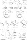
Examples of environment sensitive fluorescent labels that have been incorporated into PNA probes as a tethered label.
Figure 19
Figure 19
The mechanism of fluorescence change in TO dye.
Figure 20
Figure 20
Fluorescent nucleobases capable of hydrogen bonding that have been incorporated into PNA probes.
Figure 21
Figure 21
Comparison of the designs of the (A) light-up PNA probe and (B) FIT PNA probe.
Figure 22
Figure 22
The structures of TO and its analogues that have successfully been used in FIT PNA probes.
Figure 23
Figure 23
The working principle of dual-labeled FIT PNA probes [–223].

References

    1. Heather J M, Chain B. Genomics. 2016;107:1–8. doi: 10.1016/j.ygeno.2015.11.003. - DOI - PMC - PubMed
    1. Mascini M, Palchetti I, Tombelli S. Angew Chem, Int Ed. 2012;51:1316–1332. doi: 10.1002/anie.201006630. - DOI - PubMed
    1. Tyagi S, Kramer F R. Nat Biotechnol. 1996;14:303–308. doi: 10.1038/nbt0396-303. - DOI - PubMed
    1. Tyagi S, Bratu D P, Kramer F R. Nat Biotechnol. 1998;16:49–53. doi: 10.1038/nbt0198-49. - DOI - PubMed
    1. Nielsen P E. Acc Chem Res. 1999;32:624–630. doi: 10.1021/ar980010t. - DOI

LinkOut - more resources