. 2018 Mar 2:10:27.
doi: 10.1186/s13148-018-0462-7. eCollection 2018.

Overexpression of UHRF1 promotes silencing of tumor suppressor genes and predicts outcome in hepatoblastoma

Affiliations

Overexpression of UHRF1 promotes silencing of tumor suppressor genes and predicts outcome in hepatoblastoma

Alexander Beck et al. Clin Epigenetics. .

Abstract

Background: Hepatoblastoma (HB) is the most common liver tumor of childhood and occurs predominantly within the first 3 years of life. In accordance to its early manifestation, HB has been described to display an extremely low mutation rate. As substitute, epigenetic modifiers seem to play an exceptional role in its tumorigenesis, which holds promise to develop targeted therapies and establish biomarkers for patient risk stratification.

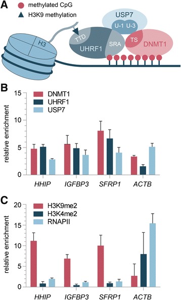
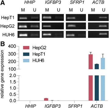
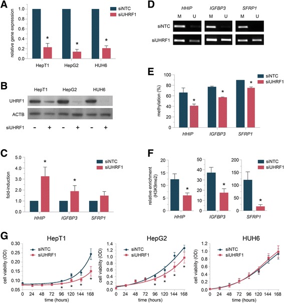
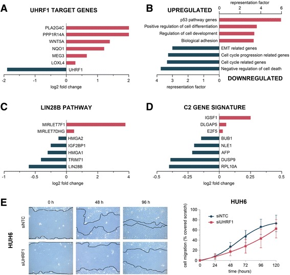
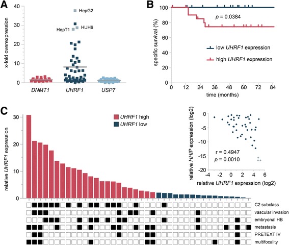
Results: We examined the role of a newly described protein complex consisting of three epigenetic regulators, namely E3 ubiquitin-like containing PHD and RING finger domain 1 (UHRF1), ubiquitin-specific-processing protease 7 (USP7), and DNA methyltransferase 1 (DNMT1), in HB. We found the complex to be located on the promoter regions of the pivotal HB-associated tumor suppressor genes (TSGs) HHIP, IGFBP3, and SFRP1 in HB cells, thereby leading to strong repression through DNA methylation and histone modifications. Consequently, knockdown of UHRF1 led to DNA demethylation and loss of the repressive H3K9me2 histone mark at the TSG loci with their subsequent transcriptional reactivation. The observed growth impairment of HB cells upon UHRF1 knockdown could be attributed to reduced expression of genes involved in cell cycle progression, negative regulation of cell death, LIN28B signaling, and the adverse 16-gene signature, as revealed by global RNA sequencing. Clinically, overexpression of UHRF1 in primary tumor tissues was significantly associated with poor survival and the prognostic high-risk 16-gene signature.

Conclusion: These findings suggest that UHRF1 is critical for aberrant TSG silencing and sustained growth signaling in HB and that UHRF1 overexpression levels might serve as a prognostic biomarker and potential molecular target for HB patients.

Keywords: Biomarker; DNA methylation; DNMT1; Epigenetic silencing; Hepatoblastoma; Histone methylation; Tumor suppressor genes; UHRF1; USP7.


Conflict of interest statement

Written informed consent was obtained from each patient, and the study protocol was approved by the Committee of Ethics of the Ludwig Maximilian University of Munich (no. 431-11).Not applicableThe authors declare that they have no competing interests.Springer Nature remains neutral with regard to jurisdictional claims in published maps and institutional affiliations.

Figures

Fig. 1
Fig. 1
a Schematic overview of the trimeric complex comprising UHRF1, USP7, and DNMT1. Involved protein interactions and protein domains according to Felle et al. are shown [11]. b, c Chromatin immunoprecipitation (ChIP) was performed with HUH6 cells and the indicated antibodies. The average of two independent ChIP experiments for b DNMT1, UHRF1, and USP7 as well as c H3K9me2, H3K4me2, and RNAPII is shown. Standard deviations, genes of interest, and antibodies used for ChIP are indicated. The enrichment of specific IP versus IgG background is plotted
Fig. 2
Fig. 2
a Methylation status (U, unmethylated; M, methylated) of the HHIP, IGFBP3, SFRP1, and ACTB promoter region was determined for the indicated cell lines by MSP. Representative images of MSP experiments are shown. b Relative expression levels of indicated genes compared to the TBP housekeeping gene in three liver tumor cell lines are given. The average of three independent experiments is shown
Fig. 3
Fig. 3
a Knockdown of UHRF1 in indicated cell lines. Relative gene expression in cells transfected with siRNA against UHRF1 compared to negative control (siNTC) after 48 h is shown. Data represents average of three independent experiments. b Western blot analysis of HepT1, HepG2, and HUH6 cells 48 h after UHRF1 knockdown. c Relative expression levels of indicated genes in HUH6 cells 48 h after UHRF1 knockdown compared to negative control. The average of three independent experiments is shown. d Methylation status (U, unmethylated; M, methylated) of the HHIP, IGFBP3, and SFRP1 promoter region 48 h after UHRF1 knockdown was determined by MSP in HUH6 cells. Representative images of MSP experiments are shown. e Pyrosequencing results of promoter regions of indicated genes in HUH6 cells 48 h after UHRF1 knockdown of three independent experiments are shown. f ChIP analyses in HUH6 cells 48 h after UHRF1 knockdown. The relative enrichment of H3K9me2 at the indicated gene promoter regions is shown. g Cell viability of HepG2, HepT1, and HUH6 cells as evaluated by MTT assay at the indicated time points after UHRF1 knockdown. Values represent means ± standard deviation of three independent experiments performed in duplicates. Statistical significance of all experiments was calculated using t test (p < 0.05)
Fig. 4
Fig. 4
RNA sequencing results upon UHRF1 knockdown in HUH6 cells. Log2 fold change of a known UHRF1 target genes, c genes related to the LIN28B pathway, and d genes highly expressed in the adverse C2 gene signature. b Significant enrichment of up- or downregulated genes within the indicated gene ontology and hallmark gene sets. Enrichment is indicated by the corresponding representation factor of each gene set. e Scratch assay. Representative images of HUH6 cells transfected with siRNA against UHRF1 migrating into a cell-free space at 0, 48, and 96 h compared to negative control (siNTC). Quantitative evaluation of scratch closure shows the mean values of two independent experiments
Fig. 5
Fig. 5
a The relative expression of DNMT1, UHRF1, and USP7 in 40 primary HB and three HB cell lines (depicted in gray) compared to the mean of seven normal liver tissues are given. b Overall survival was calculated as time from diagnosis to death of the disease and is plotted for 40 HB patients. Statistical significance was calculated using the Mantel-Cox test. c Individual UHRF1 expression levels of 40 primary HBs are shown with the occurrence of important clinicopathological characteristics depicted as black boxes below. Inset: Expression levels of UHRF1 and HHIP in primary tumor tissues (dark blue dots) and tumor cell lines (light blue dots) were plotted against each other, and Spearman’s rank correlation was performed

Similar articles

Cited by

References

    1. Zimmermann A. The emerging family of hepatoblastoma tumours: from ontogenesis to oncogenesis. Eur J Cancer. 2005;41:1503–1514. doi: 10.1016/j.ejca.2005.02.035. - DOI - PubMed
    1. Czauderna P, Lopez-Terrada D, Hiyama E, Haberle B, Malogolowkin MH, Meyers RL. Hepatoblastoma state of the art: pathology, genetics, risk stratification, and chemotherapy. Curr Opin Pediatr. 2014;26:19–28. doi: 10.1097/MOP.0000000000000046. - DOI - PubMed
    1. Meyers RL, Maibach R, Hiyama E, Haberle B, Krailo M, Rangaswami A, Aronson DC, Malogolowkin MH, Perilongo G, von Schweinitz D, et al. Risk-stratified staging in paediatric hepatoblastoma: a unified analysis from the Children’s Hepatic tumors International Collaboration. Lancet Oncol. 2017;18:122–131. doi: 10.1016/S1470-2045(16)30598-8. - DOI - PMC - PubMed
    1. Zsiros J, Maibach R, Shafford E, Brugieres L, Brock P, Czauderna P, Roebuck D, Childs M, Zimmermann A, Laithier V, et al. Successful treatment of childhood high-risk hepatoblastoma with dose-intensive multiagent chemotherapy and surgery: final results of the SIOPEL-3HR study. J Clin Oncol. 2010;28:2584–2590. doi: 10.1200/JCO.2009.22.4857. - DOI - PubMed
    1. Cairo S, Armengol C, De Reynies A, Wei Y, Thomas E, Renard CA, Goga A, Balakrishnan A, Semeraro M, Gresh L, et al. Hepatic stem-like phenotype and interplay of Wnt/beta-catenin and Myc signaling in aggressive childhood liver cancer. Cancer Cell. 2008;14:471–484. doi: 10.1016/j.ccr.2008.11.002. - DOI - PubMed

Publication types

Substances