

The effects of taxanes, vorinostat and doxorubicin on growth and proliferation of Echinococcus multilocularis metacestodes assessed with magnetic resonance imaging and simultaneous positron emission tomography
- PMID: 29507675
- PMCID: PMC5823665
- DOI: 10.18632/oncotarget.24142
The effects of taxanes, vorinostat and doxorubicin on growth and proliferation of Echinococcus multilocularis metacestodes assessed with magnetic resonance imaging and simultaneous positron emission tomography
Abstract
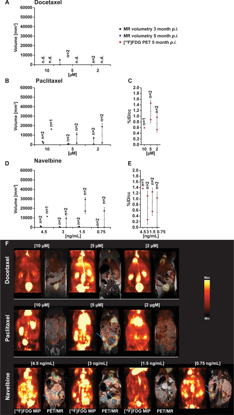
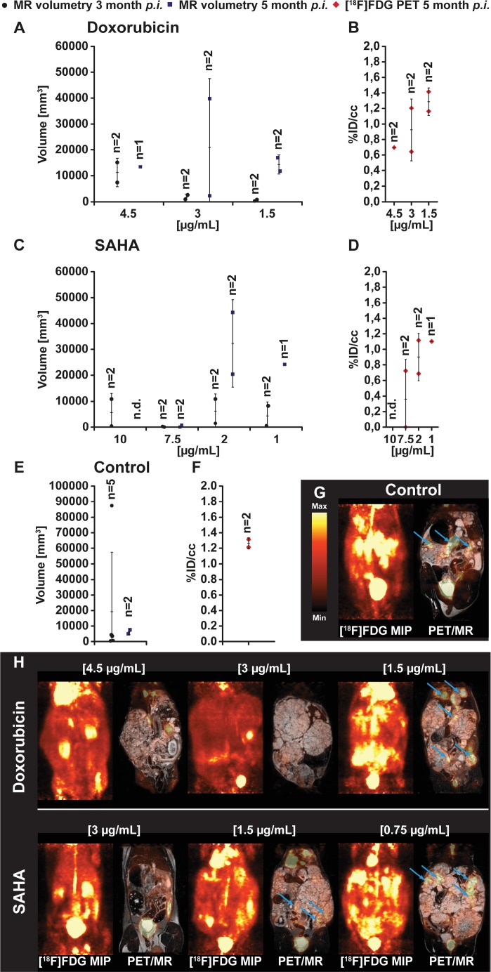
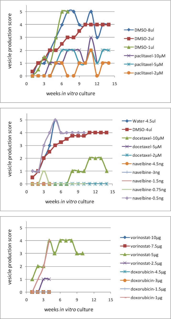
Cytostatic drugs used in cancer therapy were evaluated for their capacity to inhibit Echinococcus multilocularis metacestode growth and proliferation. Metacestode tissues were exposed in vitro to docetaxel, doxorubicin, navelbine, paclitaxel, and vorinostat for 1 week, then incubated in drug-free culture, and thereafter metacestodes were injected into the peritoneum of Meriones unguiculatus. Magnetic resonance imaging (MRI) and simultaneous positron emission tomography (PET) were applied to monitor in vivo growth of drug-exposed E. multilocularis in Meriones. At 3 month p.i., docetaxel (at 10 μM, 5 μM and 2 μM) inhibited in vivo growth and proliferation of E. multilocularis, and at 5 months p.i., only in the 2 μM docetaxel exposure group 0.3 cm 3 of parasite tissue was found. With paclitaxel and navelbine the in vivo growth of metacestodes was suppressed until 3 months p.i., thereafter, parasite tissues enlarged up to 3 cm 3 in both groups. E. multilocularis tissues of more than 10 g developed in Meriones injected with metacestodes which were previously exposed in vitro to doxorubicin, navelbine, paclitaxel or vorinostat. In Meriones infected with metacestodes previously exposed to docetaxel, the in vivo grown parasite tissues weighted 0.2 g. In vitro cultured E. multilocularis metacestodes exposed to docetaxel did not produce vesicles until 7 weeks post drug exposure, while metacestodes exposed to doxorubicin, navelbine and vorinostat proliferated continuously. In summary, docetaxel, and less efficaciously paclitaxel, inhibited in vivo and in vitro parasite growth and proliferation, and these observations suggest further experimental studies with selected drug combinations which may translate into new treatment options against alveolar echinococcosis.
Keywords: drug exposure; echinococcus multilocularis, metacestode; magnetic resonance imaging; positron emission tomography; taxanes, paclitaxel, docetaxel, histone deacetylase inhibitor, vorinostat, doxorubicin.
Conflict of interest statement
CONFLICTS OF INTEREST The authors have no conflicts of interest to disclose.
Figures

References
-
- Lubinsky G, Lee CF, Baron RW. Attempts at chemotherapy of Echinococcus multilocularis infections in rodents. II. A study of some parasiticides and cytostatic agents. Canadian journal of zoology. 1971;49:1301–1304. - PubMed
-
- Novak M. Efficacy of mitomycin C against alveolar Echinococcus. International journal of parasitology. 1990;20:119–120. - PubMed
-
- Liance M, Nemati F, Bories C, Couvreur P. Experience with doxorubicin-bound polyisohexylcyanoacrylate nanoparticles on murine alveolar echinococcosis of the liver. Int J Parasitol. 1993;23:427–9. - PubMed
-
- Naguleswaran A, Spicher M, Vonlaufen N, Ortega-Mora LM, Torgerson P, Gottstein B, Hemphill A. In vitro metacestodicidal activities of genistein and other isoflavones against Echinococcus multilocularis and Echinococcus granulosus. Antimicrobial Agents and Chemotherapy. 2006;50:3770–3778. - PMC - PubMed
LinkOut - more resources
Full Text Sources
Other Literature Sources
