Review
. 2017 Jul 6;2(4):210-220.
doi: 10.1136/svn-2017-000079. eCollection 2017 Dec.

Eyes and stroke: the visual aspects of cerebrovascular disease

Affiliations
Review

Eyes and stroke: the visual aspects of cerebrovascular disease

John H Pula et al. Stroke Vasc Neurol. .

Abstract

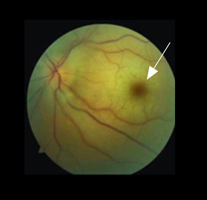
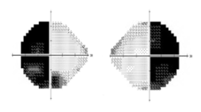
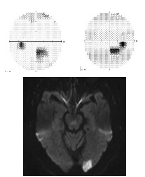
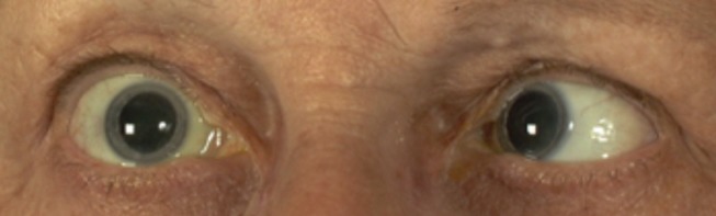
A large portion of the central nervous system is dedicated to vision and therefore strokes have a high likelihood of involving vision in some way. Vision loss can be the most disabling residual effect after a cerebral infarction. Transient vision problems can likewise be a harbinger of stroke and prompt evaluation after recognition of visual symptoms can prevent future vascular injury. In this review, we discuss the visual aspects of stroke. First, anatomy and the vascular supply of the visual system are considered. Then, the different stroke syndromes which involve vision are discussed. Finally, topics involving the assessment, prognosis, treatment and therapeutic intervention of vision-specific stroke topics are reviewed.

Keywords: Amaurosis Fugax; Diplopia; Homonymous hemianopia; Stroke; Vision.


Conflict of interest statement

Competing interests: None declared.

Figures

Figure 1
Figure 1
Radiographic angiography of the posterior circulation. Bilateral vertebral arteries fuse to form the basilar artery. The ultimate branches of the basilar artery form the posterior cerebral arteries (PCAs, arrows), which supply the visual cortex. Infarctions in the PCA territories can cause cortical vision loss. Bilateral PCA infarcts as in ‘top of the basilar syndrome’ can result in cortical blindness.
Figure 2
Figure 2
Fundus photo of central retinal artery occlusion (CRAO). CRAO results in generalised retinal oedema, sparing the optic nerve, which is supported by a separate blood supply. Examination shows a ‘cherry red spot’ (arrow). The cherry red spot is an optical illusion. The macula is not more red; the oedema of the nerve fibres surrounding it colours it pallid, causing the macula, which has relatively less nerve fibres and therefore less tissue to swell, to appear relatively red.
Figure 3
Figure 3
Humphrey visual field showing bitemporal hemianopia. Visual fields are represented with the left eye on the left and right eye on the right, which is opposite to how CT or MRI is viewed. Therefore, the temporal fields are laterally represented. Bitemporal hemianopia will generally respect the vertical meridian and imply the chiasm as the location of the insult.
Figure 4
Figure 4
Visual fields and diffusion-weighted imaging MRI showing occipital infarction. Humphrey visual fields (above) show a partial congruous right inferior field defect. Diffusion-weighted MRI (below) shows an acute infarction of the left occipital pole. The visual fields correlate in terms of localisation to the radiological defect.
Figure 5
Figure 5
Sixth nerve palsy. In this image, the patient is asked to look right. His right eye does not abduct, while his left eye adducts normally. This is typical of an abducens (sixth nerve) palsy.
Figure 6
Figure 6
Hollenhorst plaque. Fundus photo showing a chalky-coloured cholesterol plaque in the superior retinal vascular arcade.
Figure 7
Figure 7
Frisén stages of papilloedema. Five fundus photos showing the various stages of papilloedema. (A) Grade 1: Obscured nasal disc border. (B) Grade 2: Obscuration of all borders with halo. (C) Grade 3: Obscuration of a segment of blood vessel. (D) Grade 4: Total obscuration of one of the blood vessels on the disc head. (E) Grade 5: Dome-shaped protrusion with obscuration of vessels of the entire optic disc.
Figure 8
Figure 8
Terson’s syndrome. Terson’s syndrome showing a subretinal haemorrhage after an acute intracranial haemorrhage.
Figure 9
Figure 9
Ocular sequela of cavernous-carotid fistula. Right eye of a patient with a cavernous-carotid fistula showing proptosis, chemosis and conjunctival injection.
Figure 10
Figure 10
Anatomy of visual structures on MRI. (A) Coronal orbital view (arrowhead at medial rectus muscle). (B) Axial FLAIR MRI (arrow at occipital pole). (C) Axial fat-saturated orbital sequence (arrow at optic nerve).

References

    1. Rowe FJ, Wright D, Brand D, et al. . A prospective profile of visual field loss following stroke: prevalence, type, rehabilitation, and outcome. Biomed Res Int 2013;2013:1–12. (719096) doi:10.1155/2013/719096 - DOI - PMC - PubMed
    1. Sand KM, Wilhelmsen G, Naess H, et al. . Vision problems in ischaemic stroke patients: effects on life quality and disability. Eur J Neurol 2016;23:1–7. doi:10.1111/ene.12848 - DOI - PubMed
    1. Varma DD, Cugati S, Lee AW, et al. . A review of central retinal artery occlusion: clinical presentation and management. Eye 2013;27:688–97. doi:10.1038/eye.2013.25 - DOI - PMC - PubMed
    1. de Vries TM, Aries MJ, De Groot JC, et al. . A grid-like hemi-field defect following a lacunar infarct in the lateral geniculate nucleus. Clin Neurol Neurosurg 2012;114:278–80. doi:10.1016/j.clineuro.2011.10.021 - DOI - PubMed
    1. Tsuda H, Yoshizawa T. Localized infarction of the lateral geniculate body. Intern Med 2014;53:1891–2. doi:10.2169/internalmedicine.53.2695 - DOI - PubMed